01 / Morfologi

Ciri dan struktur

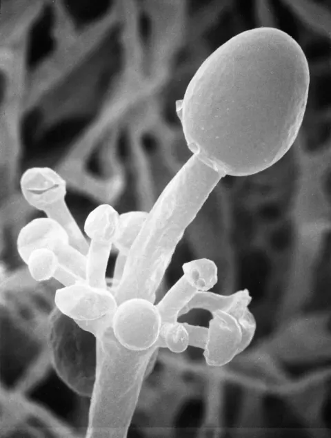
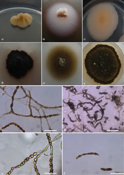
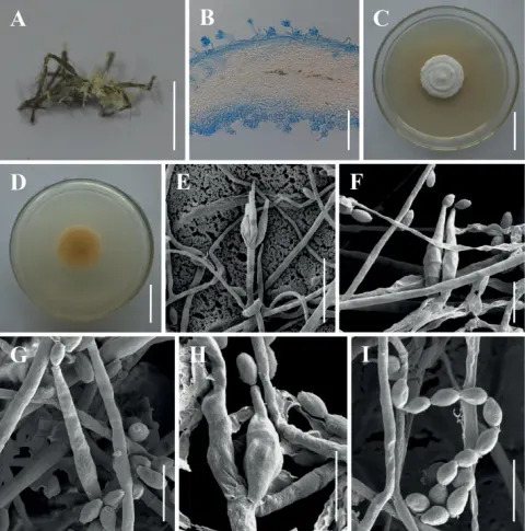
Acrophialophora fusispora is similar to Paecilomyces, but differ in the presence of pigmented, warted conidiophores, verticillate phialides in limited numbers with narrowing tip, and frequent sympodial proliferation. A. fusispora is distinguished by its pigmented fusiform conidia, which are covered by spiral bands, measuring 5–12 x 3–6 μm. The conidia arises in long single-cell basipetal chains, ranging from colorless to pale-brown and broadly ellipsoidal to lemon-shaped. The conidiophores arise singly, terminally, and laterally from the hyphae, and are erect or ascending, straight or flexuous, smooth or rough, and septate. Moreover, the conidiophores are thick-walled with brown at the base and pale towards the apex. The conidiophores are up to 1.5 μm long and 2–5 μm wide, with whorls of phialides at the end. The phialides are flask-shaped and swollen near the base, with a long, narrow neck, and hyaline, smooth or spiny wall. The phialides are borne on conidiophores, or sometimes, on vegetative hyphae, with the broadest part measuring 9–15 μm x 3–4.5 μm. Acrophialophora fusispora only reproduces asexually. The colonies of A. fusispora are dense, matted and felt-like at the base, with cottony aerial hyphae. The color of the colonies is initially dull white, but becoming grayish-brown, with black colony reverse. The colonies can reach 4–7 cm in diameter within 14 days at 25 °C on Malt Extract Agar. In vitro growth was found to be stimulated by exudates and extracts of Cassia tora. The fungus also grows well and sporulates freely on potato dextrose agar.

Sebagian uraian ditampilkan dalam bahasa sumber; terjemahannya belum tersedia atau belum lolos pemeriksaan.

Rujukan: [1]

Pengukuran mengikuti objek, spesimen atau kultur, dan metode yang disebutkan sumber. Baca uraian asli untuk konteks lengkap.

02 / Kehidupan

Habitat dan peran ekologis

Acrophialophora fusispora telah diisolasi dari tanah, udara, dan berbagai tumbuhan, seperti Cajanus, Cicer, jagung, dan Tephrosia. Jamur ini ditemukan di tanah daerah tropis hingga temperat, dan dikenal dari tanah yang tidak dibudidayakan, tanah ladang, di bawah tanaman berlumpur atau rumput, tanah hutan, dan rawa garam. With exception dari catatan dari Ceko, Singapura, Kepulauan Tuamotu, Mesir, dan Pakistan, A. fusispora hampir eksklusif dilaporkan dari India. Jamur ini telah ditemukan secara umum di permukaan akar dan rhizospheres rumput, kapas, Euphorbia species dan Carica papaya, dan pada rhizoid Funaria species. Selain itu, A. fusispora juga telah diisolasi dari cacing tanah dan hutan yang terkena air laut. Selain itu, di rhizosphere Trigonella foenum-graecum, semprotan daun dengan antibiotik subamycin meningkatkan potensi isolasi A. fusispora. Acrophialophora fusispora menunjukkan preferensi yang jelas untuk cakrawala tanah yang lebih dalam (30–45 cm) di India. Jamur ini tahan terhadap suhu, dengan suhu minimum 14 C, suhu optimal di 40 C, dan suhu maksimum 50 C.

Rujukan: [1]

Habitat atau konteks temuan
hutan
Substrat
Informasi belum tersedia pada sumber rilis ini.
Peran atau konteks
Belum dikategorikan pada rilis ini.
Temukan hubungan serupa
03 / Siklus hidup

Reproduksi

Acrophialophora fusispora is similar to Paecilomyces, but differ in the presence of pigmented, warted conidiophores, verticillate phialides in limited numbers with narrowing tip, and frequent sympodial proliferation. A. fusispora is distinguished by its pigmented fusiform conidia, which are covered by spiral bands, measuring 5–12 x 3–6 μm. The conidia arises in long single-cell basipetal chains, ranging from colorless to pale-brown and broadly ellipsoidal to lemon-shaped. The conidiophores arise singly, terminally, and laterally from the hyphae, and are erect or ascending, straight or flexuous, smooth or rough, and septate. Moreover, the conidiophores are thick-walled with brown at the base and pale towards the apex.

Sebagian uraian ditampilkan dalam bahasa sumber; terjemahannya belum tersedia atau belum lolos pemeriksaan.

Rujukan: [1]

Sinonim menurut acuan taksonomi rilis: Acrophialophora nainiana; Masoniella indica; Paecilomyces fusisporus
04 / Batas informasi

Catatan identitas dan cakupan

  • Nama diterima mengikuti Catalogue of Life Base2026-09-11; uraian mempertahankan konsep dan nama sumber aslinya.
  • Original geographic, specimen and life-cycle scope retained; source import awaits individual editorial review.
  • Impor dan terjemahan otomatis belum diperiksa editorial per profil. Struktur reproduktif yang disebutkan tidak membuktikan seluruh siklus hidup diketahui.
  • Pengukuran pada uraian mengikuti spesimen, kultur, dan metode sumber; angka tidak diperluas ke semua populasi.

Catatan distribusi dalam narasi mengikuti sumbernya. Dokumentasi spesimen atau foto mengikuti catatan sumber. Bukti wilayah Indonesia dan penilaian konservasi tidak disimpulkan dari gambar.

05 / Dokumentasi

Melihat dengan konteks

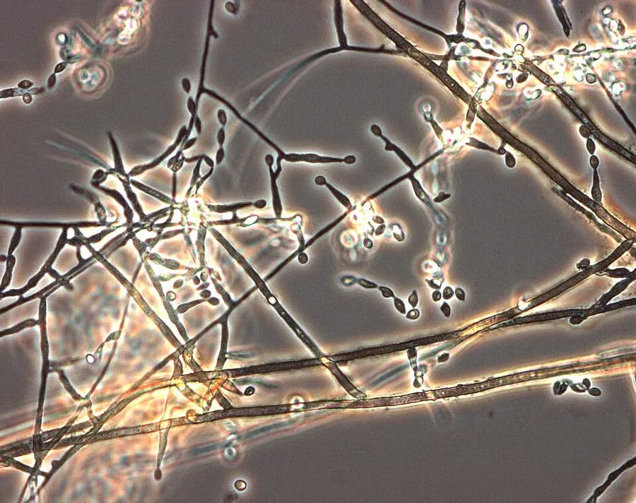
Dokumentasi Acrophialophora fusispora dari Wikimedia Commons. Acrophialophora fusispora (UAMH 11640) in phase contrast microscopy from 6 day old slide culture at 35 C Identifikasi mengikuti record berkas sumber.

Jenis
Fotografi
Kredit
Medmyco
Sumber media
Buka entri sumber

Gambar menunjukkan objek menurut identifikasi sumber, bukan verifikasi independen spesimen oleh Fungi. Tidak ada skala baru yang ditambahkan.

06 / Rujukan

Sumber profil

  1. [1] Acrophialophora fusispora — Wikipedia (en)Bagian sumber: Frozen revision 1229604583
    Tanggal penghimpunan sumber: 2026-10-12
    Teks sumber: CC BY-SA4.0 · Wikipedia contributors
  2. [2] Catalogue of Life Base2026-09-11 — Acrophialophora fusisporaBagian sumber: ColDP NameUsage.tsv ID 9NPG; accepted species; source 2073
    Tanggal penghimpunan sumber: 2026-10-12
  3. [3] Dokumentasi visual — Acrophialophora fusisporaBagian sumber: Individual media record; identification follows the source, not independent determination
    Tanggal penghimpunan sumber: 2026-10-12
    Media sumber: CC BY-SA 4.0 · Medmyco

Adaptasi teks: CC BY-SA 4.0. Wikipedia contributors — Acrophialophora fusispora — Wikipedia (en)

Uraian asli dan riwayat sumber

Riwayat atau halaman sumber

Ringkasan sumber

Acrophialophora fusispora is a poorly studied ascomycete fungus found in soil, air and various plants. A. fusispora is morphologically similar to the genera Paecilomyces and Masonia, but differ in the presence of pigmented conidiophores, verticillate phialides, and frequent sympodial proliferation. Moreover, A. fusispora is distinguished by its pigmented spindle-shaped conidia, covered with spiral bands. The fungus is naturally found in soils of tropical to temperate regions. The fungus has been identified as a plant and animal pathogen, and has recently been recognized as an emerging opportunistic human pathogen.

Revisi sumber

Morfologi sumber

Acrophialophora fusispora is similar to Paecilomyces, but differ in the presence of pigmented, warted conidiophores, verticillate phialides in limited numbers with narrowing tip, and frequent sympodial proliferation. A. fusispora is distinguished by its pigmented fusiform conidia, which are covered by spiral bands, measuring 5–12 x 3–6 μm. The conidia arises in long single-cell basipetal chains, ranging from colorless to pale-brown and broadly ellipsoidal to lemon-shaped. The conidiophores arise singly, terminally, and laterally from the hyphae, and are erect or ascending, straight or flexuous, smooth or rough, and septate. Moreover, the conidiophores are thick-walled with brown at the base and pale towards the apex. The conidiophores are up to 1.5 μm long and 2–5 μm wide, with whorls of phialides at the end. The phialides are flask-shaped and swollen near the base, with a long, narrow neck, and hyaline, smooth or spiny wall. The phialides are borne on conidiophores, or sometimes, on vegetative hyphae, with the broadest part measuring 9–15 μm x 3–4.5 μm. Acrophialophora fusispora only reproduces asexually. The colonies of A. fusispora are dense, matted and felt-like at the base, with cottony aerial hyphae. The color of the colonies is initially dull white, but becoming grayish-brown, with black colony reverse. The colonies can reach 4–7 cm in diameter within 14 days at 25 °C on Malt Extract Agar. In vitro growth was found to be stimulated by exudates and extracts of Cassia tora. The fungus also grows well and sporulates freely on potato dextrose agar.

Revisi sumber

Ekologi sumber

Acrophialophora fusispora has been isolated from soil, air, and various plants, such as Cajanus, Cicer, maize, and Tephrosia. The fungus found in the soil of tropical to temperate regions, and is known from uncultivated soils, field soils, under leguminous plants or grasses, forest soils, and salt-marshes. With exception of records from Czechoslovakia, Singapore, Tuamotu Archipelago, Egypt, and Pakistan, A. fusispora is almost exclusively reported from India. The fungus has been found commonly on root surfaces and rhizospheres of grasses, cotton, Euphorbia species and Carica papaya, and on rhizoids of Funaria species. Additionally, A. fusispora has also been isolated from earthworm casts and woods exposed to seawater. Moreover, in the rhizosphere of Trigonella foenum-graecum, foliar spray with the antibiotic subamycin increases the potential for A. fusispora isolation. Acrophialophora fusispora exhibits a distinct preference for deeper (30–45 cm) soil horizon in India. The fungus is thermotolerant, with a minimum temperature of 14 C, optimum temperature at 40 C, and maximum temperature of 50 C.

Revisi sumber

Struktur reproduksi sumber

Acrophialophora fusispora is similar to Paecilomyces, but differ in the presence of pigmented, warted conidiophores, verticillate phialides in limited numbers with narrowing tip, and frequent sympodial proliferation. A. fusispora is distinguished by its pigmented fusiform conidia, which are covered by spiral bands, measuring 5–12 x 3–6 μm. The conidia arises in long single-cell basipetal chains, ranging from colorless to pale-brown and broadly ellipsoidal to lemon-shaped. The conidiophores arise singly, terminally, and laterally from the hyphae, and are erect or ascending, straight or flexuous, smooth or rough, and septate. Moreover, the conidiophores are thick-walled with brown at the base and pale towards the apex.

Revisi sumber

Rujukan halaman ini

Fungi. Acrophialophora fusispora. Rilis data fungi-2026-10-12-expansion-2. https://fungi-atlas.mgadib.chatgpt.site/jamur/acrophialophora-fusispora/.

Status: impor sumber; pemeriksaan editorial per profil belum selesai. Untuk klaim biologis, gunakan rujukan pada bagian terkait dan periksa konteks sumber aslinya.