Ciri dan struktur
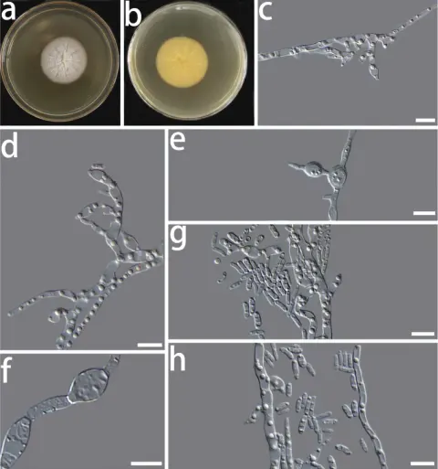
Colony on PDA after 8 d, hyphae hyaline, multi-guttulate, septate, smooth-walled; conidiophores short; conidiogenous cells hyaline, phialidic or oval in shape, single or in clusters on short lateral branches, measuring 2.9–7.2 × 1.8–3.7 μm (x̄= 4.7 × 2.6 μm, n = 50) (Fig. 2f, g); two types of conidia were observed, swollen conidia were hyaline, one-celled, dumb-bell-shaped, with hyphae emerging from both ends (Fig. 2d, e), measuring 7.6–16.5 × 2.3–4.1 μm (x̄ = 9.7 × 3.1 μm, n = 50) (Fig. 2c), oblong conidia were hyaline, one-celled, often oblong to ellipsoidal in shape, measuring 3.4–6.8 × 1.4–2.7 μm (x̄ = 4.7 × 1.8 μm, n = 50) (Fig. 2h). Chlamydospores absent. Sexual morph unknown. The optimal temperature for growth was 25 °C on PDA. No growth was detected at 5 and 35 °C. Colonies on PDA after 8 d at 25 °C were white, circular, margin entire, flat, dense, partially immersed in the medium and sticky protuberance at the centre of the colony.
Habitat dan peran ekologis
China, Yunnan Province: Tiesuo township, 26°32'71"N, 100°57'3"E, ca. 2120 m elev., isolated from Candelaria fibrosa, 13 Nov 2020, H. L. Si, CX03C1 (HMAS 350271, holotype), ex-type culture CGMCC3.20304. Colony on PDA after 8 d, hyphae hyaline, multi-guttulate, septate, smooth-walled; conidiophores short; conidiogenous cells hyaline, phialidic or oval in shape, single or in clusters on short lateral branches, measuring 2.9–7.2 × 1.8–3.7 μm (x̄= 4.7 × 2.6 μm, n = 50) (Fig. 2f, g); two types of conidia were observed, swollen conidia were hyaline, one-celled, dumb-bell-shaped, with hyphae emerging from both ends (Fig. 2d, e), measuring 7.6–16.5 × 2.3–4.1 μm (x̄ = 9.7 × 3.1 μm, n = 50) (Fig. 2c), oblong conidia were hyaline, one-celled, often oblong to ellipsoidal in shape, measuring 3.4–6.8 × 1.4–2.7 μm (x̄ = 4.7 × 1.8 μm, n = 50) (Fig. 2h). Chlamydospores absent. Sexual morph unknown. The optimal temperature for growth was 25 °C on PDA. No growth was detected at 5 and 35 °C. Colonies on PDA after 8 d at 25 °C were white, circular, margin entire, flat, dense, partially immersed in the medium and sticky protuberance at the centre of the colony. China, Yunnan Province: Tiesuo township, 26°32'71"N, 100°57'3"E, ca. 2120 m elev., isolated from on Candelaria fibrosa, 13 Nov 2020, H.
- Habitat atau konteks temuan
- kultur yang didokumentasikan
- Substrat
- Informasi belum tersedia pada sumber rilis ini.
- Peran atau konteks
- Belum dikategorikan pada rilis ini.
Reproduksi
Colony on PDA after 8 d, hyphae hyaline, multi-guttulate, septate, smooth-walled; conidiophores short; conidiogenous cells hyaline, phialidic or oval in shape, single or in clusters on short lateral branches, measuring 2.9–7.2 × 1.8–3.7 μm (x̄= 4.7 × 2.6 μm, n = 50) (Fig. 2f, g); two types of conidia were observed, swollen conidia were hyaline, one-celled, dumb-bell-shaped, with hyphae emerging from both ends (Fig. 2d, e), measuring 7.6–16.5 × 2.3–4.1 μm (x̄ = 9.7 × 3.1 μm, n = 50) (Fig. 2c), oblong conidia were hyaline, one-celled, often oblong to ellipsoidal in shape, measuring 3.4–6.8 × 1.4–2.7 μm (x̄ = 4.7 × 1.8 μm, n = 50) (Fig. 2h). Chlamydospores absent. Sexual morph unknown.
Catatan identitas dan cakupan
- Nama diterima mengikuti Catalogue of Life Base2026-09-11; uraian mempertahankan konsep dan nama sumber aslinya.
- Preserve original treatment localities, hosts and substrate scope. Materials examined describe documented specimens, not the full global range.
- Impor dan terjemahan otomatis belum diperiksa editorial per profil. Struktur reproduktif yang disebutkan tidak membuktikan seluruh siklus hidup diketahui.
- Pengukuran pada uraian mengikuti spesimen, kultur, dan metode sumber; angka tidak diperluas ke semua populasi.
Catatan distribusi dalam narasi mengikuti sumbernya. Dokumentasi spesimen atau foto mengikuti catatan sumber. Bukti wilayah Indonesia dan penilaian konservasi tidak disimpulkan dari gambar.
Melihat dengan konteks
Morphological characters of Coniochaeta fibrosae sp. nov. (HMAS 350271) a, b cultures on PDA from the surface and reverse c swollen conidia d, e swollen conidia germinate hyphae f, g conidiogenous cells h conidia. Scale bars: 10 μm.
- Jenis
- Pelat ilmiah
- Kredit
- Hong-Li Si, Yue-Min Su, Xiao-Xiao Zheng, Meng-Yao Ding, Tanay Bose, Run-Lei Chang
- Lisensi
- CC BY 4.0
- Sumber media
- Buka entri sumber
Sumber profil
- [1] Phylogenetic and morphological analyses of Coniochaeta isolates recovered from Inner Mongolia and Yunnan revealed three new endolichenic fungal species — Coniochaeta fibrosae
- [2] Catalogue of Life Base2026-09-11 — Coniochaeta fibrosae
- [3] Dokumentasi visual — Coniochaeta fibrosae
Rujukan halaman ini
Fungi. Coniochaeta fibrosae. Rilis data fungi-2026-10-12-expansion-2. https://fungi-atlas.mgadib.chatgpt.site/jamur/coniochaeta-fibrosae/.
Status: impor sumber; pemeriksaan editorial per profil belum selesai. Untuk klaim biologis, gunakan rujukan pada bagian terkait dan periksa konteks sumber aslinya.