Ciri dan struktur
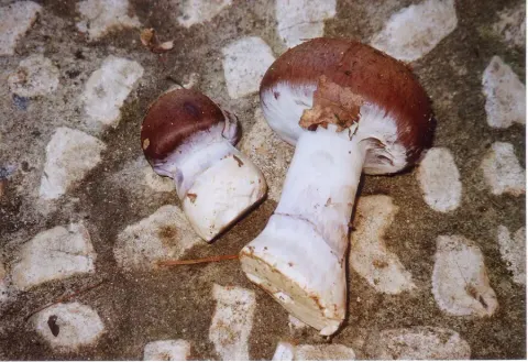
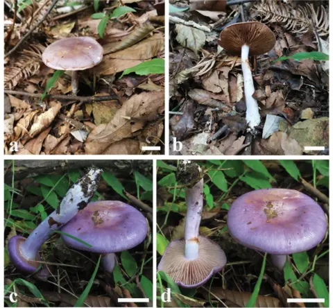
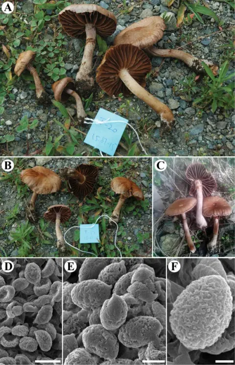
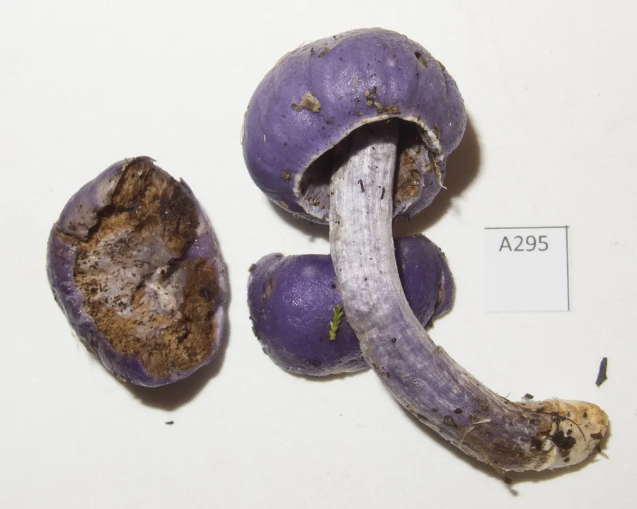
The fruitingbodies of this common fungus in New Zealand can vary considerably in shape and size as is shown in Fig. 3 a-d. Excellent descriptions have been given by Cunningham (1942: 84), Heim (1951: 129) and Singer & Smith (1958: 212) but the white veil remnants scattered along the margin of the gastrocarp or along the stipe hitherto have been overlooked. All collections kept in our herbarium showed in fresh conditions obvious veil remnants, which cover parts of the peridium and still more frequently the lower portion of the stipe. Spores 12-1.5 x 7.5-9.5 µm, ellipsoid to ellipsoid-oblong, rust brown, verrucose, mostly axially symmetric, perisporium poorly developed or lacking. Basidia 35-50 x 10-14 µm 4-spored. Cystidia absent. Epicutis consisting of interwoven, gelatinised, hyaline hyphae (2-6 µm diam.) forming a cutis, hyphae of subcutis cylindrical, with thin-walled membrane and epimembranous pigment. Clamp connections present, but sometimes rare.
Habitat dan peran ekologis
On ground amongst litter of Nothofagus fusca, N. menziesii, N. solandri, N. cliffortioides. New Zealand.
- Habitat atau konteks temuan
- Informasi belum tersedia pada sumber rilis ini.
- Substrat
- Informasi belum tersedia pada sumber rilis ini.
- Peran atau konteks
- Belum dikategorikan pada rilis ini.
Reproduksi
The fruitingbodies of this common fungus in New Zealand can vary considerably in shape and size as is shown in Fig. 3 a-d. Excellent descriptions have been given by Cunningham (1942: 84), Heim (1951: 129) and Singer & Smith (1958: 212) but the white veil remnants scattered along the margin of the gastrocarp or along the stipe hitherto have been overlooked. All collections kept in our herbarium showed in fresh conditions obvious veil remnants, which cover parts of the peridium and still more frequently the lower portion of the stipe. Spores 12-1.5 x 7.5-9.5 µm, ellipsoid to ellipsoid-oblong, rust brown, verrucose, mostly axially symmetric, perisporium poorly developed or lacking. Basidia 35-50 x 10-14 µm 4-spored.
Catatan identitas dan cakupan
- Nama diterima mengikuti Catalogue of Life Base2026-09-11; uraian mempertahankan konsep dan nama sumber aslinya.
- Historical description as cited by BiotaNZ. Preserve explicit New Zealand/locality, host and specimen qualifiers. Distribution is the cited source scope, not a current global range.
- Impor dan terjemahan otomatis belum diperiksa editorial per profil. Struktur reproduktif yang disebutkan tidak membuktikan seluruh siklus hidup diketahui.
- Pengukuran pada uraian mengikuti spesimen, kultur, dan metode sumber; angka tidak diperluas ke semua populasi.
Catatan distribusi dalam narasi mengikuti sumbernya. Dokumentasi spesimen atau foto mengikuti catatan sumber. Bukti wilayah Indonesia dan penilaian konservasi tidak disimpulkan dari gambar.
Melihat dengan konteks
Dokumentasi Cortinarius porphyroideus menurut asosiasi foto pada record takson iNaturalist. Kredit: Jerry Cooper. Identitas mengikuti sumber; bukan penentuan spesimen independen.
- Jenis
- Fotografi
- Kredit
- (c) Jerry Cooper, some rights reserved (CC BY), uploaded by Jerry Cooper
- Lisensi
- CC BY 4.0
- Sumber media
- Buka entri sumber
Dokumentasi tambahan
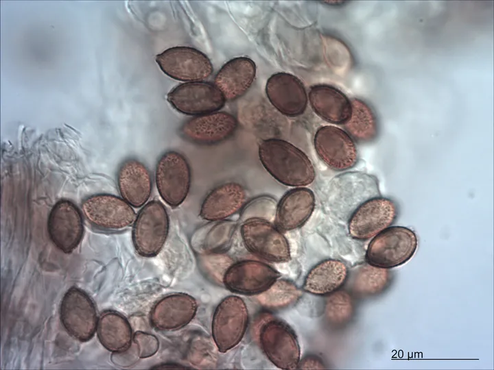
Foto yang diasosiasikan dengan Cortinarius porphyroideus pada iNaturalist. Identifikasi mengikuti sumber; bukan penentuan spesimen independen.
Kredit dan penggunaan
(c) Jerry Cooper, some rights reserved (CC BY), uploaded by Jerry Cooper
Ukuran dan format gambar disesuaikan tanpa pemotongan, retus, atau penambahan skala. Lisensi sumber dipertahankan.
Sumber profil
- [1] Horak, E. 1973: Fungi Agaricini Novazelandiae I-V. Beihefte zur Nova Hedwigia. 43. — Thaxterogaster porphyreus
- [2] Catalogue of Life Base2026-09-11 — Cortinarius porphyroideus
- [3] Dokumentasi visual — Cortinarius porphyroideus
- [4] Dokumentasi visual — Cortinarius porphyroideus
Rujukan halaman ini
Fungi. Cortinarius porphyroideus. Rilis data fungi-2026-10-12-expansion-2. https://fungi-atlas.mgadib.chatgpt.site/jamur/cortinarius-porphyroideus/.
Status: impor sumber; pemeriksaan editorial per profil belum selesai. Untuk klaim biologis, gunakan rujukan pada bagian terkait dan periksa konteks sumber aslinya.