01 / Morfologi

Ciri dan struktur

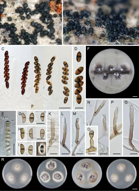
Conidiophores 200–350 μm long (x̅ = 280.6 μm, SD = 45, n = 20), 7–10 µm wide (x̅ = 8.6 μm, SD = 1.9, n = 20), mononematous, erect, simple, straight or flexuous, thick-walled, dark brown, paler towards the apex, 8–10 septate. Conidiogenous cells 48.5–60 μm long (x̅ = 53.5 μm, SD = 3.9, n = 20), 9–12 µm wide (x̅ = 10.2 μm, SD = 1.3, n = 20), with a narrow cytoplasmic channel and marked periclinal thickening in the upper quarter, lacking a collarette, proliferating enteroblastically to produce successive conidia at the same level. Conidia 21.5–40 μm long (x̅ = 29.3 μm, SD = 6.8, n = 20), 7.5–10 µm wide (x̅ = 8.2 μm, SD = 0.5, n = 20), holoblastic, solitary, accumulating in translucent slimy masses at the apices of conidiogenous, cylindrical, obtuse at the apex, slightly tapered towards the truncate base, hyaline, 3–euseptate, smooth, eguttulate. Also see Bhat and Sutton (1985) and Xia et al. (2013).

Sebagian uraian ditampilkan dalam bahasa sumber; terjemahannya belum tersedia atau belum lolos pemeriksaan.

Rujukan: [1]

Pengukuran mengikuti objek, spesimen atau kultur, dan metode yang disebutkan sumber. Baca uraian asli untuk konteks lengkap.

02 / Kehidupan

Habitat dan peran ekologis

Type status: Other material. Occurrence: recordedBy: Chun-Sheng Long; occurrenceID: C1A8F2EF-0EC1-5EDB-B7AD-6279AD758FF5; Taxon: scientificName: Kylindria excentrica; Location: continent: Asia; country: China; stateProvince: Guizhou; county: Libo county; verbatimElevation: 875 m; verbatimCoordinates: 25.1205N, 107.3634E; Identification: identifiedBy: Chun-Sheng Long, Qi-Rui Li & Jian Ma; Event: eventDate: 11/21/2021; habitat: on decaying wood; Record Level: collectionID: GMB0398 Kylindria excentrica was firstly found on rotten wood in Ethiopia (Bhat and Sutton 1985). Kylindria excentrica is similar to K. millettiae Y.D. Zhang & X.G. Zhang in morphology, but differs markedly in conidial dimensions (19.5–24 × 6.5–9 μm vs. 27.5–35 × 7.5–8.5 μm) (Zhang et al. 2010). In addition, K. excentrica has a lateral flat scar in the conidial base, whereas those of K. millettiae have a lateral flat scar in the excentric (Zhang et al. 2010).

Sebagian uraian ditampilkan dalam bahasa sumber; terjemahannya belum tersedia atau belum lolos pemeriksaan.

Rujukan: [1]

Habitat atau konteks temuan
Informasi belum tersedia pada sumber rilis ini.
Substrat
kayu
Peran atau konteks
Belum dikategorikan pada rilis ini.
Temukan hubungan serupa
03 / Siklus hidup

Reproduksi

Conidiophores 200–350 μm long (x̅ = 280.6 μm, SD = 45, n = 20), 7–10 µm wide (x̅ = 8.6 μm, SD = 1.9, n = 20), mononematous, erect, simple, straight or flexuous, thick-walled, dark brown, paler towards the apex, 8–10 septate. Conidiogenous cells 48.5–60 μm long (x̅ = 53.5 μm, SD = 3.9, n = 20), 9–12 µm wide (x̅ = 10.2 μm, SD = 1.3, n = 20), with a narrow cytoplasmic channel and marked periclinal thickening in the upper quarter, lacking a collarette, proliferating enteroblastically to produce successive conidia at the same level. Conidia 21.5–40 μm long (x̅ = 29.3 μm, SD = 6.8, n = 20), 7.5–10 µm wide (x̅ = 8.2 μm, SD = 0.5, n = 20), holoblastic, solitary, accumulating in translucent slimy masses at the apices of conidiogenous, cylindrical, obtuse at the apex, slightly tapered towards the truncate base, hyaline, 3–euseptate, smooth, eguttulate.

Sebagian uraian ditampilkan dalam bahasa sumber; terjemahannya belum tersedia atau belum lolos pemeriksaan.

Rujukan: [1]

Sinonim menurut acuan taksonomi rilis: Kylindria excentrica
04 / Batas informasi

Catatan identitas dan cakupan

  • Nama diterima mengikuti Catalogue of Life Base2026-09-11; uraian mempertahankan konsep dan nama sumber aslinya.
  • Preserve original treatment localities, hosts and substrate scope. Materials examined describe documented specimens, not the full global range.
  • Impor dan terjemahan otomatis belum diperiksa editorial per profil. Struktur reproduktif yang disebutkan tidak membuktikan seluruh siklus hidup diketahui.
  • Pengukuran pada uraian mengikuti spesimen, kultur, dan metode sumber; angka tidak diperluas ke semua populasi.

Catatan distribusi dalam narasi mengikuti sumbernya. Dokumentasi spesimen atau foto mengikuti catatan sumber. Bukti wilayah Indonesia dan penilaian konservasi tidak disimpulkan dari gambar.

05 / Dokumentasi

Melihat dengan konteks

Dokumentasi visual yang memenuhi pemeriksaan subjek dan hak penggunaan belum tersedia. Foto gejala, label, dan gambar yang meragukan tidak dihitung.

06 / Rujukan

Sumber profil

  1. [1] Additions to hyphomycetes from Yungui Plateau, China with three new species (Ascomycota, Sordariomycetes) — Kylindria excentricaBagian sumber: PMC10848564_section_53
    Tanggal penghimpunan sumber: 2026-10-12
    Teks sumber: CC BY 4.0 · Chun-Sheng L, You-Peng W, Xu Z, Yan L, Xiang-Chun S, Jian M, Qi-Rui LI.
  2. [2] Catalogue of Life Base2026-09-11 — Cylindrotrichum excentricumBagian sumber: ColDP NameUsage.tsv ID 334H6; accepted species; source 2073
    Tanggal penghimpunan sumber: 2026-10-12

Adaptasi teks: CC BY 4.0. Chun-Sheng L, You-Peng W, Xu Z, Yan L, Xiang-Chun S, Jian M, Qi-Rui LI. — Additions to hyphomycetes from Yungui Plateau, China with three new species (Ascomycota, Sordariomycetes) — Kylindria excentrica

Uraian asli dan riwayat sumber

Riwayat atau halaman sumber

Ringkasan sumber

Conidiophores 200–350 μm long (x̅ = 280.6 μm, SD = 45, n = 20), 7–10 µm wide (x̅ = 8.6 μm, SD = 1.9, n = 20), mononematous, erect, simple, straight or flexuous, thick-walled, dark brown, paler towards the apex, 8–10 septate. Conidiogenous cells 48.5–60 μm long (x̅ = 53.5 μm, SD = 3.9, n = 20), 9–12 µm wide (x̅ = 10.2 μm, SD = 1.3, n = 20), with a narrow cytoplasmic channel and marked periclinal thickening in the upper quarter, lacking a collarette, proliferating enteroblastically to produce successive conidia at the same level.

Revisi sumber

Morfologi sumber

Conidiophores 200–350 μm long (x̅ = 280.6 μm, SD = 45, n = 20), 7–10 µm wide (x̅ = 8.6 μm, SD = 1.9, n = 20), mononematous, erect, simple, straight or flexuous, thick-walled, dark brown, paler towards the apex, 8–10 septate. Conidiogenous cells 48.5–60 μm long (x̅ = 53.5 μm, SD = 3.9, n = 20), 9–12 µm wide (x̅ = 10.2 μm, SD = 1.3, n = 20), with a narrow cytoplasmic channel and marked periclinal thickening in the upper quarter, lacking a collarette, proliferating enteroblastically to produce successive conidia at the same level. Conidia 21.5–40 μm long (x̅ = 29.3 μm, SD = 6.8, n = 20), 7.5–10 µm wide (x̅ = 8.2 μm, SD = 0.5, n = 20), holoblastic, solitary, accumulating in translucent slimy masses at the apices of conidiogenous, cylindrical, obtuse at the apex, slightly tapered towards the truncate base, hyaline, 3–euseptate, smooth, eguttulate. Also see Bhat and Sutton (1985) and Xia et al. (2013).

Revisi sumber

Ekologi sumber

Type status: Other material. Occurrence: recordedBy: Chun-Sheng Long; occurrenceID: C1A8F2EF-0EC1-5EDB-B7AD-6279AD758FF5; Taxon: scientificName: Kylindria excentrica; Location: continent: Asia; country: China; stateProvince: Guizhou; county: Libo county; verbatimElevation: 875 m; verbatimCoordinates: 25.1205N, 107.3634E; Identification: identifiedBy: Chun-Sheng Long, Qi-Rui Li & Jian Ma; Event: eventDate: 11/21/2021; habitat: on decaying wood; Record Level: collectionID: GMB0398 Kylindria excentrica was firstly found on rotten wood in Ethiopia (Bhat and Sutton 1985). Kylindria excentrica is similar to K. millettiae Y.D. Zhang & X.G. Zhang in morphology, but differs markedly in conidial dimensions (19.5–24 × 6.5–9 μm vs. 27.5–35 × 7.5–8.5 μm) (Zhang et al. 2010). In addition, K. excentrica has a lateral flat scar in the conidial base, whereas those of K. millettiae have a lateral flat scar in the excentric (Zhang et al. 2010).

Revisi sumber

Struktur reproduksi sumber

Conidiophores 200–350 μm long (x̅ = 280.6 μm, SD = 45, n = 20), 7–10 µm wide (x̅ = 8.6 μm, SD = 1.9, n = 20), mononematous, erect, simple, straight or flexuous, thick-walled, dark brown, paler towards the apex, 8–10 septate. Conidiogenous cells 48.5–60 μm long (x̅ = 53.5 μm, SD = 3.9, n = 20), 9–12 µm wide (x̅ = 10.2 μm, SD = 1.3, n = 20), with a narrow cytoplasmic channel and marked periclinal thickening in the upper quarter, lacking a collarette, proliferating enteroblastically to produce successive conidia at the same level. Conidia 21.5–40 μm long (x̅ = 29.3 μm, SD = 6.8, n = 20), 7.5–10 µm wide (x̅ = 8.2 μm, SD = 0.5, n = 20), holoblastic, solitary, accumulating in translucent slimy masses at the apices of conidiogenous, cylindrical, obtuse at the apex, slightly tapered towards the truncate base, hyaline, 3–euseptate, smooth, eguttulate.

Revisi sumber

Rujukan halaman ini

Fungi. Cylindrotrichum excentricum. Rilis data fungi-2026-10-12-expansion-2. https://fungi-atlas.mgadib.chatgpt.site/jamur/cylindrotrichum-excentricum/.

Status: impor sumber; pemeriksaan editorial per profil belum selesai. Untuk klaim biologis, gunakan rujukan pada bagian terkait dan periksa konteks sumber aslinya.