Ciri dan struktur
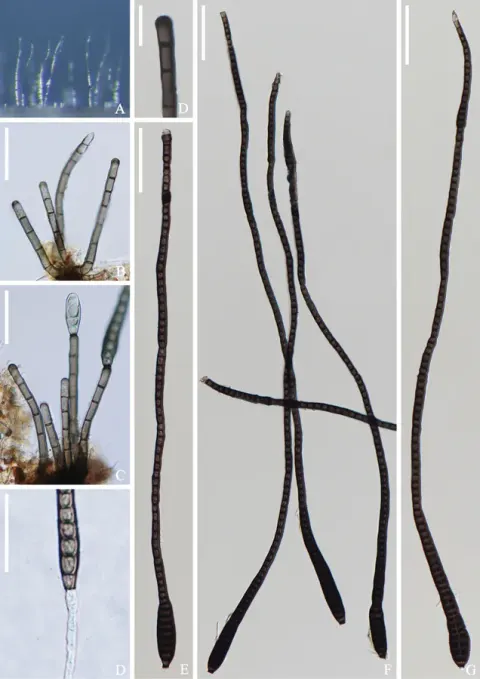
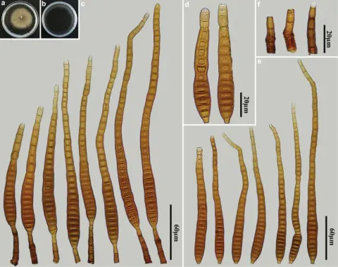
Saprobic on culms of bamboo. Sexual morph: Undetermined. Asexual morph: Hyphomycetous (Figs 1, 2). Colonies effuse, brown to dark-brown, hairy. Mycelium mostly immersed, composed of pale to dark brown, septate, branched, smooth, hyaline to subhyaline hyphae. Conidiophores macronematous, mononematous, septate, single or in groups of 2 or 3, erect, cylindrical, straight or slightly flexuous, olivaceous or brown, robust at the base 40–96 × 4–5.5 μm (x̅ = 69 × 5 μm, n = 10). Conidiogenous cells blastic, integrated, terminal, cylindrical, olivaceous or brown 9–19 × 4–5 μm (x̅ = 15 × 4.5 μm, n = 15). Conidia acrogenous, solitary, straight, obclavate, septate, thick-walled, rounded at the apex, truncate at the base, tapering towards apex, olivaceous or brown, 45–74 μm long (x̅ = 60.5 μm, n = 20), 5.5–9.5 μm at the widest (x̅ = 7.5 μm, n = 20). Culture characteristics: Conidia germinated on PDA within 12 hours and germ tubes were produced from both ends. Colony reached 30 mm in 4 weeks at 26℃ on PDA media, circular, flat, surface rough, grey from above, brown from below, edge entire.
Habitat dan peran ekologis
Saprobic on culms of bamboo. Sexual morph: Undetermined. Asexual morph: Hyphomycetous (Figs 1, 2). Colonies effuse, brown to dark-brown, hairy. Mycelium mostly immersed, composed of pale to dark brown, septate, branched, smooth, hyaline to subhyaline hyphae. Conidiophores macronematous, mononematous, septate, single or in groups of 2 or 3, erect, cylindrical, straight or slightly flexuous, olivaceous or brown, robust at the base 40–96 × 4–5.5 μm (x̅ = 69 × 5 μm, n = 10). Conidiogenous cells blastic, integrated, terminal, cylindrical, olivaceous or brown 9–19 × 4–5 μm (x̅ = 15 × 4.5 μm, n = 15). Conidia acrogenous, solitary, straight, obclavate, septate, thick-walled, rounded at the apex, truncate at the base, tapering towards apex, olivaceous or brown, 45–74 μm long (x̅ = 60.5 μm, n = 20), 5.5–9.5 μm at the widest (x̅ = 7.5 μm, n = 20). Culture characteristics: Conidia germinated on PDA within 12 hours and germ tubes were produced from both ends. Colony reached 30 mm in 4 weeks at 26℃ on PDA media, circular, flat, surface rough, grey from above, brown from below, edge entire. The morphological characteristics of Distoseptispora bambusae match well with the generic concept of Distoseptispora (Su et al. 2016).
- Habitat atau konteks temuan
- kultur yang didokumentasikan
- Substrat
- Informasi belum tersedia pada sumber rilis ini.
- Peran atau konteks
- Pengurai
Reproduksi
Saprobic on culms of bamboo. Sexual morph: Undetermined. Asexual morph: Hyphomycetous (Figs 1, 2). Colonies effuse, brown to dark-brown, hairy. Mycelium mostly immersed, composed of pale to dark brown, septate, branched, smooth, hyaline to subhyaline hyphae. Conidiophores macronematous, mononematous, septate, single or in groups of 2 or 3, erect, cylindrical, straight or slightly flexuous, olivaceous or brown, robust at the base 40–96 × 4–5.5 μm (x̅ = 69 × 5 μm, n = 10). Conidiogenous cells blastic, integrated, terminal, cylindrical, olivaceous or brown 9–19 × 4–5 μm (x̅ = 15 × 4.5 μm, n = 15). Conidia acrogenous, solitary, straight, obclavate, septate, thick-walled, rounded at the apex, truncate at the base, tapering towards apex, olivaceous or brown, 45–74 μm long (x̅ = 60.5 μm, n = 20), 5.5–9.5 μm at the widest (x̅ = 7.5 μm, n = 20).
Catatan identitas dan cakupan
- Nama diterima mengikuti Catalogue of Life Base2026-09-11; uraian mempertahankan konsep dan nama sumber aslinya.
- Preserve original treatment localities, hosts and substrate scope. Materials examined describe documented specimens, not the full global range.
- Impor dan terjemahan otomatis belum diperiksa editorial per profil. Struktur reproduktif yang disebutkan tidak membuktikan seluruh siklus hidup diketahui.
- Pengukuran pada uraian mengikuti spesimen, kultur, dan metode sumber; angka tidak diperluas ke semua populasi.
Catatan distribusi dalam narasi mengikuti sumbernya. Dokumentasi spesimen atau foto mengikuti catatan sumber. Bukti wilayah Indonesia dan penilaian konservasi tidak disimpulkan dari gambar.
Melihat dengan konteks
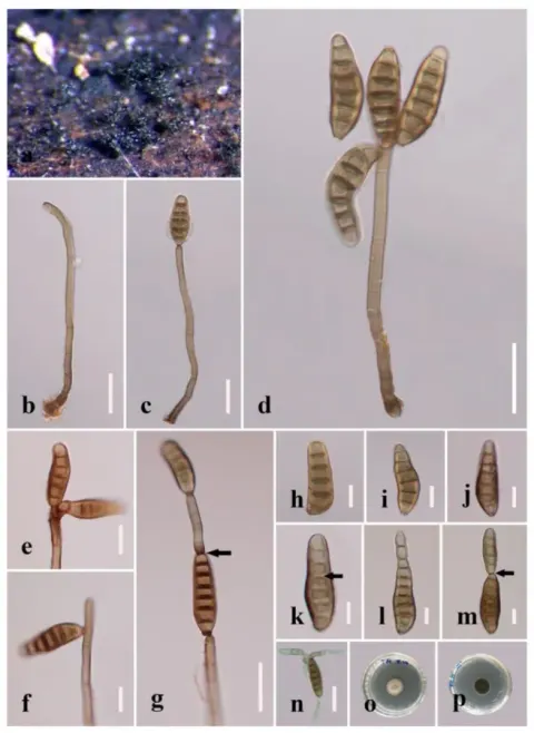
Distoseptispora bambusae (HKAS 125826) a, b colonies on woody substrates c–e conidiophores f, g conidiogenous cells h–m conidia n germinated conidium o culture on PDA. Scale bars: 50 μm (c–e); 10 μm (f, g); 20 μm (h–n).
- Jenis
- Pelat ilmiah
- Kredit
- Hong-Wei Shen, Dan-Feng Bao, Saranyaphat Boonmee, Yong-Zhong Lu, Xi-Jun Su, Yun-Xia Li, Zong-Long Luo
- Lisensi
- CC BY 4.0
- Sumber media
- Buka entri sumber
Dokumentasi tambahan

Distoseptispora bambusae. (a) Colonies on natural substrate. (b – d) Conidiophores with conidia. (e – k) Conidia. (l) Colony on PDA (from front). (m) Colony on PDA (from reverse). Scale bars: (b – d) 30 μm; (e – k) 20 μm.
Kredit dan penggunaan
© 2022 by the authors.
Ukuran dan format gambar disesuaikan tanpa pemotongan, retus, atau penambahan skala. Lisensi sumber dipertahankan.
Sumber profil
- [1] Distoseptispora bambusae sp. nov. (Distoseptisporaceae) on bamboo from China and Thailand — Distoseptispora bambusae
- [2] Catalogue of Life Base2026-09-11 — Distoseptispora bambusae
- [3] Dokumentasi visual — Distoseptispora bambusae
- [4] Dokumentasi visual — Distoseptispora bambusae
Rujukan halaman ini
Fungi. Distoseptispora bambusae. Rilis data fungi-2026-10-12-expansion-2. https://fungi-atlas.mgadib.chatgpt.site/jamur/distoseptispora-bambusae/.
Status: impor sumber; pemeriksaan editorial per profil belum selesai. Untuk klaim biologis, gunakan rujukan pada bagian terkait dan periksa konteks sumber aslinya.