Ciri dan struktur
Spider hosts covered by light yellow to pale yellow (158A–B) mycelium. Sexual morph: Stromata stipitate, solitary or multiple. Stipes cylindrical, arising from the dorsal region of the host, white to pale yellow, 3–5 mm long, 0.5–1 mm broad. Fertile heads produce at the terminal of stipes, disc-shaped, upper surface slightly convex, 1–3 × 1–2 mm. Perithecia completely immersed, narrowly ovoid, 500–750 × 200–300 µm, ostioles strong orange yellow (163B). Asci cylindrical, 8-spored, 350–450 µm long, 5–7 µm broad, with cap 3–5 µm thick. Ascospores hyaline, filiform, whole ascospores, 400–460 × 1–1.5 µm. Asexual morph: Synnemata multiple, cylindrical, occasionally acuminate apex, white, up to 8 mm long, 50–200 µm broad. Conidiogenous cells phialidic, scattered along with the synnemata. Phialides solitary, globose to subglobose, arising from the mycelium, (4)5–5.5(6) × (4)5–5.5(6) µm, with distinct necks, 0.5–1.5 × 0.5–1 µm. Conidia hyaline, fusoid or fusiform-elliptical, (2)6–8(10) × 1–2(2.5) µm. Colonies on OA attaining a diam. of 18–20 mm in 21 days, cottony with high mycelium density in the middle of colonies, mycelium with low density around the margin of colonies, flattened, white, reverse deep pink (180D). Sparse synnemata with conidiogenous cells producing conidia observed after 30 days, white, on the edge of a colonies. Phialides solitary, globose to subglobose, (4)5.5–6.5(7) × 3.5–5(5.5) µm, distinct necks, 1–3 × 0.5–1 µm. Conidia hyaline, fusoid, fusiform-elliptical, (2)6–10(13) × 1–3 µm. Colonies on PDA attaining a diam. of 7–9(10) mm in 21 days, cottony with high mycelium density, white, moderate purplish-red to dark purplish-pink (186B–C) pigment diffusing in the medium, reverse moderate red (180 A–B).
Habitat dan peran ekologis
Colonies on OA attaining a diam. of 18–20 mm in 21 days, cottony with high mycelium density in the middle of colonies, mycelium with low density around the margin of colonies, flattened, white, reverse deep pink (180D). Sparse synnemata with conidiogenous cells producing conidia observed after 30 days, white, on the edge of a colonies. Phialides solitary, globose to subglobose, (4)5.5–6.5(7) × 3.5–5(5.5) µm, distinct necks, 1–3 × 0.5–1 µm. Conidia hyaline, fusoid, fusiform-elliptical, (2)6–10(13) × 1–3 µm. Colonies on PDA attaining a diam. of 7–9(10) mm in 21 days, cottony with high mycelium density, white, moderate purplish-red to dark purplish-pink (186B–C) pigment diffusing in the medium, reverse moderate red (180 A–B). Sporulation observed after 30 days with absence of synnemata. Phialides arising from aerial hyphae, solitary, mostly globose to subglobose, occasionally cylindrical, (4)5.5–11.5(15) × 2–3.5(5) µm, distinct necks, 0.5–2 × 0.5–1 µm. Conidia hyaline, fusoid, fusiform-elliptical, cylindrical, (2)6–9.5(11) × 1–3 µm. Specimens were found on the underside of dicot leaves of forest plants. Thailand, Nakhon Ratchasima Province, Khao Yai National Park, 14°26'20.72"N, 101°22'20.02"E, on spider (Web builder, Araneae) attached to the underside of a dicot leaf of forest plants, 10 June 2010, K.
- Habitat atau konteks temuan
- hutan; kultur yang didokumentasikan
- Substrat
- Informasi belum tersedia pada sumber rilis ini.
- Peran atau konteks
- Belum dikategorikan pada rilis ini.
Reproduksi
Spider hosts covered by light yellow to pale yellow (158A–B) mycelium. Sexual morph: Stromata stipitate, solitary or multiple. Stipes cylindrical, arising from the dorsal region of the host, white to pale yellow, 3–5 mm long, 0.5–1 mm broad. Fertile heads produce at the terminal of stipes, disc-shaped, upper surface slightly convex, 1–3 × 1–2 mm. Perithecia completely immersed, narrowly ovoid, 500–750 × 200–300 µm, ostioles strong orange yellow (163B). Asci cylindrical, 8-spored, 350–450 µm long, 5–7 µm broad, with cap 3–5 µm thick. Ascospores hyaline, filiform, whole ascospores, 400–460 × 1–1.5 µm. Asexual morph: Synnemata multiple, cylindrical, occasionally acuminate apex, white, up to 8 mm long, 50–200 µm broad.
Catatan identitas dan cakupan
- Nama diterima mengikuti Catalogue of Life Base2026-09-11; uraian mempertahankan konsep dan nama sumber aslinya.
- Preserve original treatment localities, hosts and substrate scope. Materials examined describe documented specimens, not the full global range.
- Impor dan terjemahan otomatis belum diperiksa editorial per profil. Struktur reproduktif yang disebutkan tidak membuktikan seluruh siklus hidup diketahui.
- Pengukuran pada uraian mengikuti spesimen, kultur, dan metode sumber; angka tidak diperluas ke semua populasi.
Catatan distribusi dalam narasi mengikuti sumbernya. Dokumentasi spesimen atau foto mengikuti catatan sumber. Bukti wilayah Indonesia dan penilaian konservasi tidak disimpulkan dari gambar.
Melihat dengan konteks
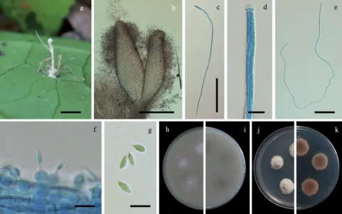
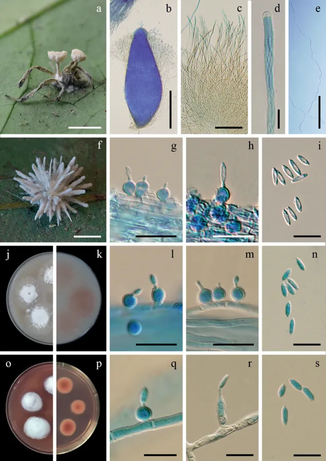
Hevansia novoguineensis a fungus on a spider (BBH 32171) b perithecium c asci d ascus tip e filiform, whole ascospore f fungus on a spider (BBH 31299) g–i phialides with conidia on synnema j, k colonies on OA at 21 days (j obverse, k reverse) l–n phialides with conidia on OA o, p colonies on PDA at 21 days with purplish-red pigment diffusing in agar medium (o obverse, p reverse) q–s phialides with conidia on PDA. Scale bars: 5 mm (a, f); 200 µm (b); 100 µm (c, e); 10 µm (d, g–i, l–n, q, r, s).
- Jenis
- Pelat ilmiah
- Kredit
- Suchada Mongkolsamrit, Wasana Noisripoom, Kanoksri Tasanathai, Noppol Kobmoo, Donnaya Thanakitpipattana, Artit Khonsanit, Booppa Petcharad, Baramee Sakolrak, Winanda Himaman
- Lisensi
- CC BY 4.0
- Sumber media
- Buka entri sumber
Sumber profil
- [1] Comprehensive treatise of Hevansia and three new genera Jenniferia, Parahevansia and Polystromomyces on spiders in Cordycipitaceae from Thailand — Hevansia novoguineensis
- [2] Catalogue of Life Base2026-09-11 — Hevansia novoguineensis
- [3] Dokumentasi visual — Hevansia novoguineensis
Rujukan halaman ini
Fungi. Hevansia novoguineensis. Rilis data fungi-2026-10-12-expansion-2. https://fungi-atlas.mgadib.chatgpt.site/jamur/hevansia-novoguineensis/.
Status: impor sumber; pemeriksaan editorial per profil belum selesai. Untuk klaim biologis, gunakan rujukan pada bagian terkait dan periksa konteks sumber aslinya.