Ciri dan struktur
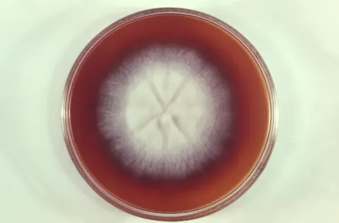
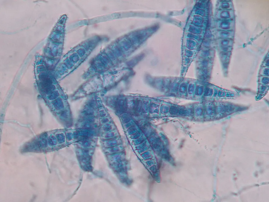
Microsporum canis forms a white, coarsely fluffy spreading colony with a distinctive "hairy" or "feathery" texture. On the underside of the growth medium, a characteristic deep yellow pigment develops due to the metabolites secreted by the fungus. The intensity of this yellow pigmentation peak on the sixth day of colony growth and fades gradually making the identification of older colonies difficult. Some strains of M. canis fail to produce yellow pigment altogether, exhibit abnormally slow colony growth and form undeveloped macroconidia. Cultivation on polished rice tends to reestablish the typical growth morphology and is helpful for identification. Microsporum canis reproduces asexually by forming macroconidia that are asymmetrical, spindle-shaped and have cell walls that are thick and coarsely roughened. The interior portion of each macroconidium is typically divided into six or more compartments separated by broad cross-walls. Microsporum canis also produces microconidia that resemble those of many other dermatophytes and thus are not a useful diagnostic feature. Typically, infections caused by M. canis are associated with alopecia in the case of tinea capitis, while ringworm infections in pets produce characteristic inflammatory lesions, which may or may not result in hair loss. This species has a propensity to cause subclinical infections in some animal species, particularly long-haired cats are frequent reservoirs of infection. Isolation of the fungus from brushed pet hair can aid in detection of either an actively growing fungus or a passive carriage of fungal hyphae or arthroconidia.
Habitat dan peran ekologis
Despite its nama spesies ("canis" berarti anjing), tuan rumah alami M. canis adalah kucing domestik. However this spesies dapat mengkolonisasi anjing dan kuda juga. Dalam semua kasus, itu tinggal di kulit dan bulu. Microsporum canis juga dapat bertahan sebagai spora yang tidak aktif di lingkungan untuk jangka waktu yang lama. Microsporum canis spesies memiliki distribusi di seluruh dunia. Extremely high kejadian telah dilaporkan di Iran, sementara insiden yang lebih rendah dikaitkan dengan England and Scandinavian countries, serta Selatan American countries. Microsporum canis jarang terjadi di beberapa bagian AS dan Eropa, dan benar-benar tidak ada di Afrika khatulistiwa.
- Habitat atau konteks temuan
- Informasi belum tersedia pada sumber rilis ini.
- Substrat
- Informasi belum tersedia pada sumber rilis ini.
- Peran atau konteks
- Belum dikategorikan pada rilis ini.
Reproduksi
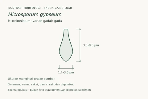
Microsporum canis reproduces asexually by forming macroconidia that are asymmetrical, spindle-shaped and have cell walls that are thick and coarsely roughened. The interior portion of each macroconidium is typically divided into six or more compartments separated by broad cross-walls. Microsporum canis also produces microconidia that resemble those of many other dermatophytes and thus are not a useful diagnostic feature.
Catatan identitas dan cakupan
- Nama diterima mengikuti Catalogue of Life Base2026-09-11; uraian mempertahankan konsep dan nama sumber aslinya.
- Original geographic, specimen and life-cycle scope retained; source import awaits individual editorial review.
- Impor dan terjemahan otomatis belum diperiksa editorial per profil. Struktur reproduktif yang disebutkan tidak membuktikan seluruh siklus hidup diketahui.
- Pengukuran pada uraian mengikuti spesimen, kultur, dan metode sumber; angka tidak diperluas ke semua populasi.
Catatan distribusi dalam narasi mengikuti sumbernya. Dokumentasi spesimen atau foto mengikuti catatan sumber. Bukti wilayah Indonesia dan penilaian konservasi tidak disimpulkan dari gambar.
Melihat dengan konteks
Dokumentasi Microsporum canis dari Wikimedia Commons. 40x Macroconidia de Microsporum canis Identifikasi mengikuti record berkas sumber.
- Jenis
- Fotografi
- Kredit
- Roberto J. Galindo
- Lisensi
- CC BY-SA 3.0
- Sumber media
- Buka entri sumber
Sumber profil
- [1] Microsporum canis — Wikipedia (en)
- [2] Catalogue of Life Base2026-09-11 — Microsporum canis
- [3] Dokumentasi visual — Microsporum canis
Rujukan halaman ini
Fungi. Microsporum canis. Rilis data fungi-2026-10-12-expansion-2. https://fungi-atlas.mgadib.chatgpt.site/jamur/microsporum-canis/.
Status: impor sumber; pemeriksaan editorial per profil belum selesai. Untuk klaim biologis, gunakan rujukan pada bagian terkait dan periksa konteks sumber aslinya.