Saprobic on batang mati Dipsacus L. Morph seksual Ascomata 200–250 × 300–350 μm (\(\bar{x} \) = 220 × 330 μm, n = 5), ditutupi oleh peridium inang, dangkal pada korteks inang, dangkal, sepi, berkeliaran, uni-lokulasi, subglobosa ke globosa tertekan, dasar datar, coklat coklat hingga coklat, dinding halus, papilate. Ostiole central, 60 × 150 μm, cokelat sampai cokelat terang, papilate, pori terbuka, ostiolate yang dipenuhi dengan perifuse. Peridium 13–40 μm lebar, tipis, seragam, terdiri dari lapisan 4–6 sel coklat terang sampai coklat tekstura angularis, lapisan dalam yang terdiri dari sel tipis, hialin.
Profil ini diimpor dari uraian spesies pada sumber berlisensi dan sebagian teks diterjemahkan otomatis. Pemeriksaan editorial per profil belum selesai. Bagian yang belum diterjemahkan atau belum lolos pemeriksaan terjemahan tetap ditampilkan dalam bahasa sumber. Angka dan nama ilmiah dipertahankan.
Saprobic on dead stems of Dipsacus L. Sexual morph Ascomata 200–250 × 300–350 μm (\(\bar{x} \) = 220 × 330 μm, n = 5), covered by host peridium, superficial on the cortex of host, superficial, solitary, scattered, uni-loculate, subglobose to depressed globose, flatten base, ligth brown to brown, smooth-walled, papillate. Ostiole central, 60 × 150 μm, brown to light brown, papillate, opened pore, ostiolate filled with periphyses. Peridium 13–40 μm wide, thin, uniform, comprising of 4–6 layers of light brown to brown cells of textura angularis, inner layers comprising thin, hyaline cells. Hamathecium of dense, 1.5–3.0 μm wide (\(\bar{x} \) = 2.5 μm, n = 50), filiform branches, anastomosing above asci, reaching the ostiole part, transverse septate, cellular pseudoparaphyses. Asci 80–120 × 10–15 μm (\(\bar{x} \) = 95 × 15 μm, n = 20), 8-spored, bitunicate, fisitunicate, thick-walled, clavate, apically rounded, with long, furcated pedicel, ocular chamber clearly visible when immature. Ascospores 18–25 × 5–8 μm (\(\bar{x} \) = 20 × 6.5 μm, n = 50), 2-seriate, partial overlapping, hyaline when immature, reddish brown to brown at maturity, cell above median septum slightly wider than below, tapering towards the ends, slightly acute at both ends, with (1–)3 transversely septa, constricted at the septa, indentation present, without mucilaginous sheath. Asexual morph Undetermined. Culture characteristics: Colonies on MEA, reaching 30 mm diam. after 4 weeks of incubation at 16 °C. Culture above cream with orange brown at the edge, medium dense mycelium, colonies circular, flat, umbonate, dull, covered with white aerial mycelium; reverse dark brown, dense, irregular, fimbriate margin.
Sebagian uraian ditampilkan dalam bahasa sumber; terjemahannya belum tersedia atau belum lolos pemeriksaan.
Pengukuran mengikuti objek, spesimen atau kultur, dan metode yang disebutkan sumber. Baca uraian asli untuk konteks lengkap.
02 / Kehidupan
Habitat dan peran ekologis
Saprobic on dead stems of Dipsacus L. Sexual morph Ascomata 200–250 × 300–350 μm (\(\bar{x} \) = 220 × 330 μm, n = 5), covered by host peridium, superficial on the cortex of host, superficial, solitary, scattered, uni-loculate, subglobose to depressed globose, flatten base, ligth brown to brown, smooth-walled, papillate. Ostiole central, 60 × 150 μm, brown to light brown, papillate, opened pore, ostiolate filled with periphyses. Peridium 13–40 μm wide, thin, uniform, comprising of 4–6 layers of light brown to brown cells of textura angularis, inner layers comprising thin, hyaline cells. Hamathecium of dense, 1.5–3.0 μm wide (\(\bar{x} \) = 2.5 μm, n = 50), filiform branches, anastomosing above asci, reaching the ostiole part, transverse septate, cellular pseudoparaphyses. Asci 80–120 × 10–15 μm (\(\bar{x} \) = 95 × 15 μm, n = 20), 8-spored, bitunicate, fisitunicate, thick-walled, clavate, apically rounded, with long, furcated pedicel, ocular chamber clearly visible when immature. Ascospores 18–25 × 5–8 μm (\(\bar{x} \) = 20 × 6.5 μm, n = 50), 2-seriate, partial overlapping, hyaline when immature, reddish brown to brown at maturity, cell above median septum slightly wider than below, tapering towards the ends, slightly acute at both ends, with (1–)3 transversely septa, constricted at the septa, indentation present, without mucilaginous sheath.
Sebagian uraian ditampilkan dalam bahasa sumber; terjemahannya belum tersedia atau belum lolos pemeriksaan.
Saprobic on dead stems of Dipsacus L. Sexual morph Ascomata 200–250 × 300–350 μm (\(\bar{x} \) = 220 × 330 μm, n = 5), covered by host peridium, superficial on the cortex of host, superficial, solitary, scattered, uni-loculate, subglobose to depressed globose, flatten base, ligth brown to brown, smooth-walled, papillate. Ostiole central, 60 × 150 μm, brown to light brown, papillate, opened pore, ostiolate filled with periphyses. Peridium 13–40 μm wide, thin, uniform, comprising of 4–6 layers of light brown to brown cells of textura angularis, inner layers comprising thin, hyaline cells. Hamathecium of dense, 1.5–3.0 μm wide (\(\bar{x} \) = 2.5 μm, n = 50), filiform branches, anastomosing above asci, reaching the ostiole part, transverse septate, cellular pseudoparaphyses.
Sebagian uraian ditampilkan dalam bahasa sumber; terjemahannya belum tersedia atau belum lolos pemeriksaan.
Nama diterima mengikuti Catalogue of Life Base2026-09-11; uraian mempertahankan konsep dan nama sumber aslinya.
Preserve original monograph treatment localities, hosts and specimen/substrate scope. Materials examined document specimens, not the full global range.
Impor dan terjemahan otomatis belum diperiksa editorial per profil. Struktur reproduktif yang disebutkan tidak membuktikan seluruh siklus hidup diketahui.
Pengukuran pada uraian mengikuti spesimen, kultur, dan metode sumber; angka tidak diperluas ke semua populasi.
Catatan distribusi dalam narasi mengikuti sumbernya. Dokumentasi spesimen atau foto mengikuti catatan sumber. Bukti wilayah Indonesia dan penilaian konservasi tidak disimpulkan dari gambar.
05 / Dokumentasi
Melihat dengan konteks
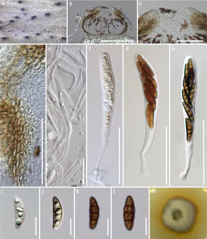
Montagnula camporesii (MFLU 16-2290, holotype). a Appearance of ascomata on Dipsacus sp. b Vertical section through ascoma. c Ostiolar canal. d Section of partial peridium layer. e Pseudoparaphyses. f – h Developmental state of asci. i – l Developmental state of ascospores. m Culture characteristic on MEA. Scale bars: b = 200 µm, c, d, f – h = 50 µm, e = 20 µm, i – l = 10 µm
Jenis
Pelat ilmiah
Kredit
Penulis dan sumber artikel: Hyde, Kevin D.; Dong, Yang; Phookamsak, Rungtiwa; Jeewon, Rajesh; Bhat, D. Jayarama; Jones, E. B. Gareth; Liu, Ning-Guo; Abeywickrama, Pranami D.; Mapook, Ausana; Wei, Deping; Perera, Rekhani H.; Manawasinghe, Ishara S.; Pem, Dhandevi; Bundhun, Digvijayini; Karunarathna, Anuruddha; Ekanayaka, Anusha H.; Bao, Dan-Feng; Li, Junfu; Samarakoon, Milan C.; Chaiwan, Napalai; Lin, Chuan-Gen; Phutthacharoen, Kunthida; Zhang, Sheng-Nan; Senanayake, Indunil C.; Goonasekara, Ishani D.; Thambugala, Kasun M.; Phukhamsakda, Chayanard; Tennakoon, Danushka S.; Jiang, Hong-Bo; Yang, Jing; Zeng, Ming; Huanraluek, Naruemon; Liu, Jian-Kui (Jack); Wijesinghe, Subodini N.; Tian, Qing; Tibpromma, Saowaluck; Brahmanage, Rashika S.; Boonmee, Saranyaphat; Huang, Shi-Ke; Thiyagaraja, Vinodhini; Lu, Yong-Zhong; Jayawardena, Ruvishika S.; Dong, Wei; Yang, Er-Fu; Singh, Sanjay K.; Singh, Shiv Mohan; Rana, Shiwali; Lad, Sneha S.; Anand, Garima; Devadatha, Bandarupalli; Niranjan, M.; Sarma, V. Venkateswara; Liimatainen, Kare; Aguirre-Hudson, Begoña; Niskanen, Tuula; Overall, Andy; Alvarenga, Renato Lúcio Mendes; Gibertoni, Tatiana Baptista; Pfliegler, Walter P.; Horváth, Enikő; Imre, Alexandra; Alves, Amanda Lucia; da Silva Santos, Ana Carla; Tiago, Patricia Vieira; Bulgakov, Timur S.; Wanasinghe, Dhanushaka N.; Bahkali, Ali H.; Doilom, Mingkwan; Elgorban, Abdallah M.; Maharachchikumbura, Sajeewa S. N.; Rajeshkumar, Kunhiraman C.; Haelewaters, Danny; Mortimer, Peter E.; Zhao, Qi; Lumyong, Saisamorn; Xu, Jianchu; Sheng, Jun
Saprobic on dead stems of Dipsacus L.
Sexual morph Ascomata 200–250 × 300–350 μm (\(\bar{x} \) = 220 × 330 μm, n = 5), covered by host peridium, superficial on the cortex of host, superficial, solitary, scattered, uni-loculate, subglobose to depressed globose, flatten base, ligth brown to brown, smooth-walled, papillate.
Ostiole central, 60 × 150 μm, brown to light brown, papillate, opened pore, ostiolate filled with periphyses.
Peridium 13–40 μm wide, thin, uniform, comprising of 4–6 layers of light brown to brown cells of textura angularis, inner layers comprising thin, hyaline cells.
Saprobic on dead stems of Dipsacus L.
Sexual morph Ascomata 200–250 × 300–350 μm (\(\bar{x} \) = 220 × 330 μm, n = 5), covered by host peridium, superficial on the cortex of host, superficial, solitary, scattered, uni-loculate, subglobose to depressed globose, flatten base, ligth brown to brown, smooth-walled, papillate.
Ostiole central, 60 × 150 μm, brown to light brown, papillate, opened pore, ostiolate filled with periphyses.
Peridium 13–40 μm wide, thin, uniform, comprising of 4–6 layers of light brown to brown cells of textura angularis, inner layers comprising thin, hyaline cells.
Hamathecium of dense, 1.5–3.0 μm wide (\(\bar{x} \) = 2.5 μm, n = 50), filiform branches, anastomosing above asci, reaching the ostiole part, transverse septate, cellular pseudoparaphyses.
Asci 80–120 × 10–15 μm (\(\bar{x} \) = 95 × 15 μm, n = 20), 8-spored, bitunicate, fisitunicate, thick-walled, clavate, apically rounded, with long, furcated pedicel, ocular chamber clearly visible when immature.
Ascospores 18–25 × 5–8 μm (\(\bar{x} \) = 20 × 6.5 μm, n = 50), 2-seriate, partial overlapping, hyaline when immature, reddish brown to brown at maturity, cell above median septum slightly wider than below, tapering towards the ends, slightly acute at both ends, with (1–)3 transversely septa, constricted at the septa, indentation present, without mucilaginous sheath.
Asexual morph Undetermined.
Culture characteristics: Colonies on MEA, reaching 30 mm diam. after 4 weeks of incubation at 16 °C.
Culture above cream with orange brown at the edge, medium dense mycelium, colonies circular, flat, umbonate, dull, covered with white aerial mycelium; reverse dark brown, dense, irregular, fimbriate margin.
Saprobic on dead stems of Dipsacus L.
Sexual morph Ascomata 200–250 × 300–350 μm (\(\bar{x} \) = 220 × 330 μm, n = 5), covered by host peridium, superficial on the cortex of host, superficial, solitary, scattered, uni-loculate, subglobose to depressed globose, flatten base, ligth brown to brown, smooth-walled, papillate.
Ostiole central, 60 × 150 μm, brown to light brown, papillate, opened pore, ostiolate filled with periphyses.
Peridium 13–40 μm wide, thin, uniform, comprising of 4–6 layers of light brown to brown cells of textura angularis, inner layers comprising thin, hyaline cells.
Hamathecium of dense, 1.5–3.0 μm wide (\(\bar{x} \) = 2.5 μm, n = 50), filiform branches, anastomosing above asci, reaching the ostiole part, transverse septate, cellular pseudoparaphyses.
Asci 80–120 × 10–15 μm (\(\bar{x} \) = 95 × 15 μm, n = 20), 8-spored, bitunicate, fisitunicate, thick-walled, clavate, apically rounded, with long, furcated pedicel, ocular chamber clearly visible when immature.
Ascospores 18–25 × 5–8 μm (\(\bar{x} \) = 20 × 6.5 μm, n = 50), 2-seriate, partial overlapping, hyaline when immature, reddish brown to brown at maturity, cell above median septum slightly wider than below, tapering towards the ends, slightly acute at both ends, with (1–)3 transversely septa, constricted at the septa, indentation present, without mucilaginous sheath.
Saprobic on dead stems of Dipsacus L.
Sexual morph Ascomata 200–250 × 300–350 μm (\(\bar{x} \) = 220 × 330 μm, n = 5), covered by host peridium, superficial on the cortex of host, superficial, solitary, scattered, uni-loculate, subglobose to depressed globose, flatten base, ligth brown to brown, smooth-walled, papillate.
Ostiole central, 60 × 150 μm, brown to light brown, papillate, opened pore, ostiolate filled with periphyses.
Peridium 13–40 μm wide, thin, uniform, comprising of 4–6 layers of light brown to brown cells of textura angularis, inner layers comprising thin, hyaline cells.
Hamathecium of dense, 1.5–3.0 μm wide (\(\bar{x} \) = 2.5 μm, n = 50), filiform branches, anastomosing above asci, reaching the ostiole part, transverse septate, cellular pseudoparaphyses.
Status: impor sumber; pemeriksaan editorial per profil belum selesai. Untuk klaim biologis, gunakan rujukan pada bagian terkait dan periksa konteks sumber aslinya.
Phylogenetic analysis berdasarkan ITS, ITS dan tub2 dan ITS dan tef1-α dataset yang dipertimbangkan dalam penelitian ini mengelompokkan strain yang dihimpun dalam garis keturunan monofiletik dalam keluarga Didymosphaeriaceae. Oleh karena…
Saprobic on batang mati dari Citrus maxima (Rutaceae). Morph seksual: tidak ditentukan. Morph asexual: Coelomycetous. konidiomata 100–150 × 100–170 µm (x̄ = 105 × 135 µm, n = 10), pycnidial, terendam pada semi-merendam, cokelat gelap…
Saprobic on batang mati dari Quercus cerris. Morph seksual tidak ditentukan. Transformasi aseksual Coelomycetous. konidiomata 300–400 μm diam., 180–250 μm tinggi, pycnidial, kesepian untuk gregarious, tenggelam ke semi-merendam, erumpent…