01 / Morfologi

Ciri dan struktur

The cap is initially convex, but as it matures, it expands to a bell-shape, typically reaching 0.3 to 1 cm in diameter. The spores are roughly spindle-shaped (i.e., tapering at each end), with dimensions of 9–11 by 3.5–4.5 μm. They are nonamyloid, meaning they do not take up iodine when stained with Melzer's reagent. The spore-bearing cells, the basidia, are club-shaped, four-spored and measure 20–22 by 5–6 μm. The cheilocystidium and pleurocystidia (cystidia found on the edge and face, respectively, of a gill) are similar, club-shaped to spindle-shaped or egg-shaped, and have apices that are often covered with a resinous secretion. The hyphae that comprise the cap cuticle are up to 3.5 μm wide, clamped, and covered with cylindrical excrescences that measure 2–9 by 1–3 μm. The hyphae of the cortical layer of the stem are up to 4.5 μm wide, clamped, and densely covered with simple to somewhat branched, cylindrical to inflated excrescences that are up to 20 by 5 μm. These latter excrescences are embedded in gelatinous matter.

Sebagian uraian ditampilkan dalam bahasa sumber; terjemahannya belum tersedia atau belum lolos pemeriksaan.

Rujukan: [1]

Pengukuran mengikuti objek, spesimen atau kultur, dan metode yang disebutkan sumber. Baca uraian asli untuk konteks lengkap.

02 / Kehidupan

Habitat dan peran ekologis

Tubuh buah tumbuh secara terpisah, dalam kelompok, atau agak berkelompok di puing-puing di tempat basah, terutama di sepanjang sungai atau perbatasan rawa. The appearance of the fruit bodies is not significantly influenced by the effect of rainfall, perhaps because "such minute fungi are largely determined by the microenvironment prevailing under dense vegetation, etc., which is no doubt less affected by recent rain than more exposed situations." The fungus is widely distributed throughout the eastern United States and Canada and occurs in Washington, Oregon, and California along the Pacific Coast. Hal ini juga dilaporkan dari Trinidad, Inggris, Norwegia, Spanyol, Korea, dan Sungai Ussuri Valley in di timur laut China.

Sebagian uraian ditampilkan dalam bahasa sumber; terjemahannya belum tersedia atau belum lolos pemeriksaan.

Rujukan: [1]

Habitat atau konteks temuan
Informasi belum tersedia pada sumber rilis ini.
Substrat
Informasi belum tersedia pada sumber rilis ini.
Peran atau konteks
Belum dikategorikan pada rilis ini.
Temukan hubungan serupa
03 / Siklus hidup

Reproduksi

The spores are roughly spindle-shaped (i.e., tapering at each end), with dimensions of 9–11 by 3.5–4.5 μm. They are nonamyloid, meaning they do not take up iodine when stained with Melzer's reagent. The spore-bearing cells, the basidia, are club-shaped, four-spored and measure 20–22 by 5–6 μm. The cheilocystidium and pleurocystidia (cystidia found on the edge and face, respectively, of a gill) are similar, club-shaped to spindle-shaped or egg-shaped, and have apices that are often covered with a resinous secretion. The hyphae that comprise the cap cuticle are up to 3.5 μm wide, clamped, and covered with cylindrical excrescences that measure 2–9 by 1–3 μm. The hyphae of the cortical layer of the stem are up to 4.5 μm wide, clamped, and densely covered with simple to somewhat branched, cylindrical to inflated excrescences that are up to 20 by 5 μm.

Sebagian uraian ditampilkan dalam bahasa sumber; terjemahannya belum tersedia atau belum lolos pemeriksaan.

Rujukan: [1]

Sinonim menurut acuan taksonomi rilis: Agaricus acicula; Hemimycena acicula; Hygrocybe acicula; Marasmiellus acicula; Mycena acicula var. longispora; Trogia acicula
04 / Batas informasi

Catatan identitas dan cakupan

  • Nama diterima mengikuti Catalogue of Life Base2026-09-11; uraian mempertahankan konsep dan nama sumber aslinya.
  • Original geographic, specimen and life-cycle scope retained; source import awaits individual editorial review.
  • Impor dan terjemahan otomatis belum diperiksa editorial per profil. Struktur reproduktif yang disebutkan tidak membuktikan seluruh siklus hidup diketahui.
  • Pengukuran pada uraian mengikuti spesimen, kultur, dan metode sumber; angka tidak diperluas ke semua populasi.

Catatan distribusi dalam narasi mengikuti sumbernya. Dokumentasi spesimen atau foto mengikuti catatan sumber. Bukti wilayah Indonesia dan penilaian konservasi tidak disimpulkan dari gambar.

05 / Dokumentasi

Melihat dengan konteks

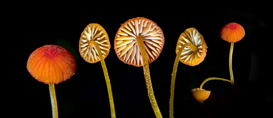
Dokumentasi Mycena acicula dari Wikimedia Commons. From https://mushroomobserver.org/273272 Identifikasi mengikuti record berkas sumber.

Jenis
Fotografi
Kredit
Alan Rockefeller
Sumber media
Buka entri sumber

Gambar menunjukkan objek menurut identifikasi sumber, bukan verifikasi independen spesimen oleh Fungi. Tidak ada skala baru yang ditambahkan.

06 / Rujukan

Sumber profil

  1. [1] Mycena acicula — Wikipedia (en)Bagian sumber: Frozen revision 1371286321
    Tanggal penghimpunan sumber: 2026-10-12
    Teks sumber: CC BY-SA4.0 · Wikipedia contributors
  2. [2] Catalogue of Life Base2026-09-11 — Mycena aciculaBagian sumber: ColDP NameUsage.tsv ID 44SMJ; accepted species; source 2073
    Tanggal penghimpunan sumber: 2026-10-12
  3. [3] Dokumentasi visual — Mycena aciculaBagian sumber: Individual media record; identification follows the source, not independent determination
    Tanggal penghimpunan sumber: 2026-10-12
    Media sumber: CC BY-SA 4.0 · Alan Rockefeller

Adaptasi teks: CC BY-SA 4.0. Wikipedia contributors — Mycena acicula — Wikipedia (en)

Uraian asli dan riwayat sumber

Riwayat atau halaman sumber

Ringkasan sumber

Mycena acicula, commonly known as the orange bonnet, or the coral spring Mycena, is a species of fungus in the family Mycenaceae. The mushrooms have small orange-red caps, up to 1 cm in diameter, held by slender yellowish stems up to 6 cm long. The gills are pale yellow with a whitish edge. Several other Mycena species look similar, but may be distinguished by differences in size and/or microscopic characteristics. The fungus grows on dead twigs and other woody debris of forest floors, especially along streams and other wet places.

Revisi sumber

Morfologi sumber

The cap is initially convex, but as it matures, it expands to a bell-shape, typically reaching 0.3 to 1 cm in diameter. The spores are roughly spindle-shaped (i.e., tapering at each end), with dimensions of 9–11 by 3.5–4.5 μm. They are nonamyloid, meaning they do not take up iodine when stained with Melzer's reagent. The spore-bearing cells, the basidia, are club-shaped, four-spored and measure 20–22 by 5–6 μm. The cheilocystidium and pleurocystidia (cystidia found on the edge and face, respectively, of a gill) are similar, club-shaped to spindle-shaped or egg-shaped, and have apices that are often covered with a resinous secretion. The hyphae that comprise the cap cuticle are up to 3.5 μm wide, clamped, and covered with cylindrical excrescences that measure 2–9 by 1–3 μm. The hyphae of the cortical layer of the stem are up to 4.5 μm wide, clamped, and densely covered with simple to somewhat branched, cylindrical to inflated excrescences that are up to 20 by 5 μm. These latter excrescences are embedded in gelatinous matter.

Revisi sumber

Ekologi sumber

The fruit bodies grow singly, in groups, or somewhat clustered on debris in wet places, especially along streams or the borders of swamps. The appearance of the fruit bodies is not significantly influenced by the effect of rainfall, perhaps because "such minute fungi are largely determined by the microenvironment prevailing under dense vegetation, etc., which is no doubt less affected by recent rain than more exposed situations." The fungus is widely distributed throughout the eastern United States and Canada and occurs in Washington, Oregon, and California along the Pacific Coast. It has also been reported from Trinidad, Britain, Norway, Spain, Korea, and the Ussuri River Valley in the northeast of China.

Revisi sumber

Struktur reproduksi sumber

The spores are roughly spindle-shaped (i.e., tapering at each end), with dimensions of 9–11 by 3.5–4.5 μm. They are nonamyloid, meaning they do not take up iodine when stained with Melzer's reagent. The spore-bearing cells, the basidia, are club-shaped, four-spored and measure 20–22 by 5–6 μm. The cheilocystidium and pleurocystidia (cystidia found on the edge and face, respectively, of a gill) are similar, club-shaped to spindle-shaped or egg-shaped, and have apices that are often covered with a resinous secretion. The hyphae that comprise the cap cuticle are up to 3.5 μm wide, clamped, and covered with cylindrical excrescences that measure 2–9 by 1–3 μm. The hyphae of the cortical layer of the stem are up to 4.5 μm wide, clamped, and densely covered with simple to somewhat branched, cylindrical to inflated excrescences that are up to 20 by 5 μm.

Revisi sumber

Rujukan halaman ini

Fungi. Mycena acicula. Rilis data fungi-2026-10-12-expansion-2. https://fungi-atlas.mgadib.chatgpt.site/jamur/mycena-acicula/.

Status: impor sumber; pemeriksaan editorial per profil belum selesai. Untuk klaim biologis, gunakan rujukan pada bagian terkait dan periksa konteks sumber aslinya.