Ciri dan struktur
Sexual morph: Ascomata 225–300 μm (n = 5), on wood, perithecial, solitary, semi-immersed, unilocular, subglobose to globose, black, ostiolate, with ostiolar neck erumpent through bark of host when mature. Neck 445–645 × 35–45 μm (x̄ = 530 × 40 μm, n = 5), centrally ostiolate, contorted, lined with hyaline periphyses. Peridium 17–40 μm diam., membranous, composed of dark brown to hyaline cells of textura angularis. Hamathecium composed of 2–6 μm wide, hyaline, septate paraphyses, slightly constricted at septa and gradually narrowed towards apex. Asci 11–20 × 3–6 μm (x̄ = 15.5 × 5 μm, n = 30), 8-spored, unitunicate, clavate, with short pedicel, apically rounded. Ascospores 3–5 × 1.5–3 μm (x̄ = 3.5 × 2 μm, n = 50), bi-seriate, hyaline, oval to ellipsoid, aseptate, smooth-walled, rounded at the ends. Asexual morph: Mycelium on culture, partly superficial, composed of septate, branched, hyaline, rarely verrucose, hyphae 1.5–3 μm diam., rarely with adelophialides. Conidiophores usually arising from hyaline hyphae, mononematous, unbranched, occasionally constricted at basal septum, hyaline. Phialides 8–15 × 2–4 μm (x̄ = 9.5 × 3 μm, n = 20), terminal, monophialidic, elongate-ampulliform and attenuated at base. Conidia 2.5–6 × 1–2.5 µm (x̄ = 4 × 2 μm, n = 30), hyaline, ellipsoid to ovoid, aseptate. Ascospore germinating on PDA within 1 week at 23°C, germ tubes produced from ends. Colonies growing on PDA, reaching 2 cm diam. and sporulating after 30 days. Colonies semi-immersed to superficial, irregular in shape, flat, slightly raised, with undulate edge, slightly rough on surface, cottony to fairly fluffy, colony from above, greyish-brown (5F3–5, Ridgway 1912) at the margin, initially write to cream (5A1–3) in the centre, becoming dark brown (5F7–8) at the margin, orange-white (5B1–3) at the centre; from below, initially, greyish-brown at the margin, white at the centre, becoming dark brown at the margin, orange-white at the centre, producing brown pigmentation in agar.
Habitat dan peran ekologis
CHINA, Yunnan Province, Baoshan, stream along the roadside; saprobic on dead wood, 21 December 2016; Huang S.K. (KUN HKAS99550, holotype; MFLU MFLU18-1076, isotype); ex-type living culture (KUMCC 17-0145; KUMCC 18-0018). GenBank no. (ITS: MH399732, TUB: MH395327, ACT: MH395325; ITS: MH399733, TUB: MH395328, ACT: MH395326) Sexual morph: Ascomata 225–300 μm (n = 5), on wood, perithecial, solitary, semi-immersed, unilocular, subglobose to globose, black, ostiolate, with ostiolar neck erumpent through bark of host when mature. Neck 445–645 × 35–45 μm (x̄ = 530 × 40 μm, n = 5), centrally ostiolate, contorted, lined with hyaline periphyses. Peridium 17–40 μm diam., membranous, composed of dark brown to hyaline cells of textura angularis. Hamathecium composed of 2–6 μm wide, hyaline, septate paraphyses, slightly constricted at septa and gradually narrowed towards apex. Asci 11–20 × 3–6 μm (x̄ = 15.5 × 5 μm, n = 30), 8-spored, unitunicate, clavate, with short pedicel, apically rounded. Ascospores 3–5 × 1.5–3 μm (x̄ = 3.5 × 2 μm, n = 50), bi-seriate, hyaline, oval to ellipsoid, aseptate, smooth-walled, rounded at the ends. Asexual morph: Mycelium on culture, partly superficial, composed of septate, branched, hyaline, rarely verrucose, hyphae 1.5–3 μm diam., rarely with adelophialides.
- Habitat atau konteks temuan
- kultur yang didokumentasikan
- Substrat
- kayu
- Peran atau konteks
- Pengurai
Reproduksi
Sexual morph: Ascomata 225–300 μm (n = 5), on wood, perithecial, solitary, semi-immersed, unilocular, subglobose to globose, black, ostiolate, with ostiolar neck erumpent through bark of host when mature. Neck 445–645 × 35–45 μm (x̄ = 530 × 40 μm, n = 5), centrally ostiolate, contorted, lined with hyaline periphyses. Peridium 17–40 μm diam., membranous, composed of dark brown to hyaline cells of textura angularis. Hamathecium composed of 2–6 μm wide, hyaline, septate paraphyses, slightly constricted at septa and gradually narrowed towards apex. Asci 11–20 × 3–6 μm (x̄ = 15.5 × 5 μm, n = 30), 8-spored, unitunicate, clavate, with short pedicel, apically rounded. Ascospores 3–5 × 1.5–3 μm (x̄ = 3.5 × 2 μm, n = 50), bi-seriate, hyaline, oval to ellipsoid, aseptate, smooth-walled, rounded at the ends.
Catatan identitas dan cakupan
- Nama diterima mengikuti Catalogue of Life Base2026-09-11; uraian mempertahankan konsep dan nama sumber aslinya.
- Preserve original treatment localities, hosts and substrate scope. Materials examined describe documented specimens, not the full global range.
- Impor dan terjemahan otomatis belum diperiksa editorial per profil. Struktur reproduktif yang disebutkan tidak membuktikan seluruh siklus hidup diketahui.
- Pengukuran pada uraian mengikuti spesimen, kultur, dan metode sumber; angka tidak diperluas ke semua populasi.
Catatan distribusi dalam narasi mengikuti sumbernya. Dokumentasi spesimen atau foto mengikuti catatan sumber. Bukti wilayah Indonesia dan penilaian konservasi tidak disimpulkan dari gambar.
Melihat dengan konteks
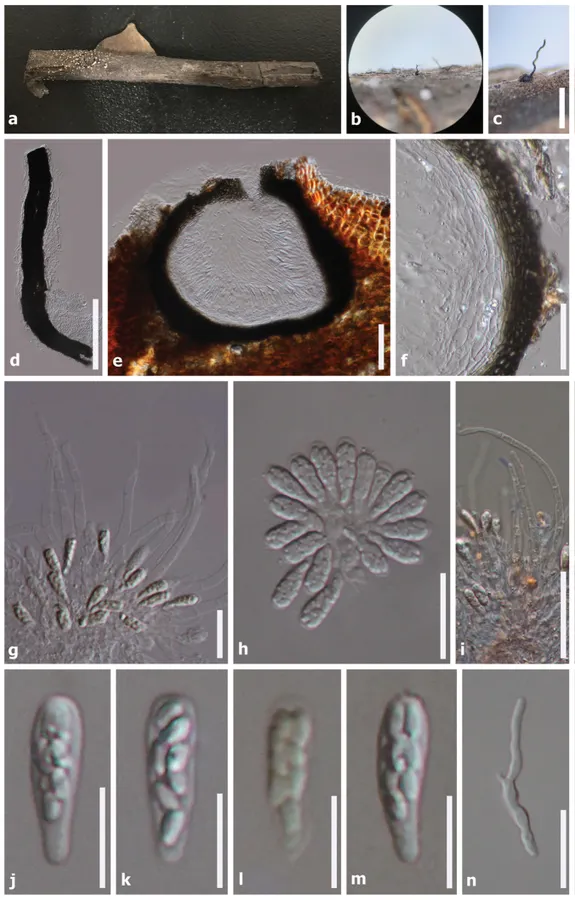
Phaeoacremonium ovale (HKAS99550, holotype). a Substrate b, c Ascoma on host d Squashed neck e Ascoma in vertical section f Peridium g Asci surrounded by paraphyses h Asci i Septate paraphyses j–m Asci with ascospores n Germinating ascospores. Note: Fig. i stained in Congo red reagent, fig l stained in Melzer’s reagent. Scale bars: 500 µm (c); 200 µm (d); 100 µm (e); 50 µm (f, i); 30 µm (n); 20 µm (g–h); 10 µm (j–m)
- Jenis
- Pelat ilmiah
- Kredit
- Shi-Ke Huang, Rajesh Jeewon, Kevin D. Hyde, D. Jayarama Bhat, Putarak Chomnunti, Ting-Chi Wen
- Lisensi
- CC BY 4.0
- Sumber media
- Buka entri sumber
Dokumentasi tambahan
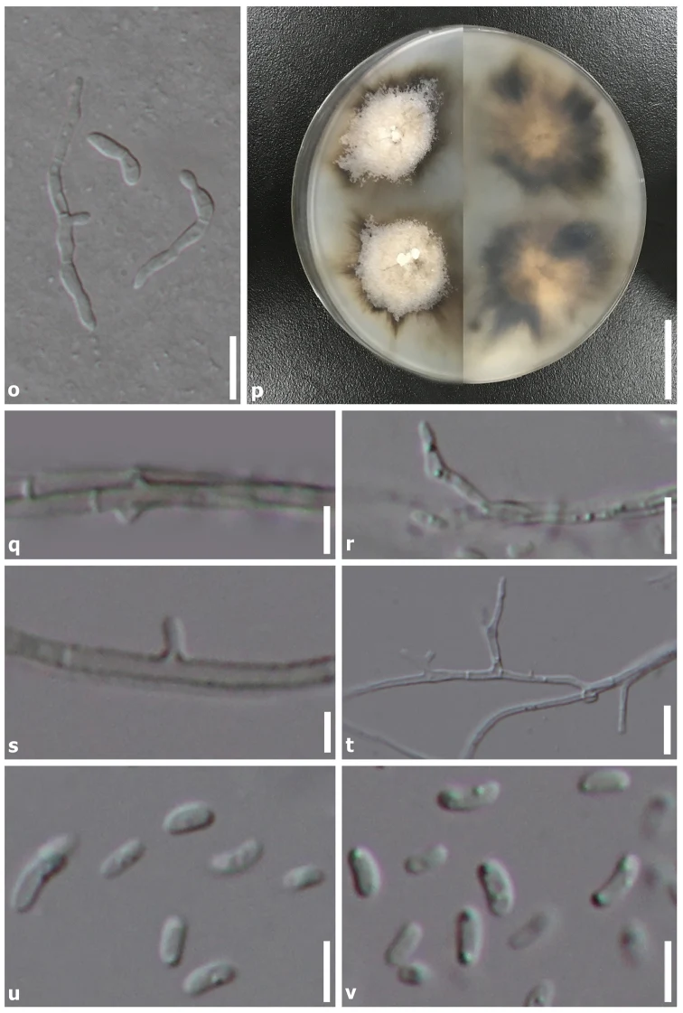
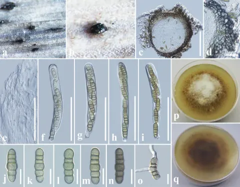
Phaeoacremonium ovale (HKAS99550, holotype). o Germinating ascospores, p 7 weeks of culture plate (above, left/reverse, right), q Mycelium with adelophialides r–t Branched conidiophores u–v Conidia. Scale bars: 20 mm (p); 20 µm (o); 10 µm (r, t); 5 µm (q, s, u–v).
Kredit dan penggunaan
Shi-Ke Huang, Rajesh Jeewon, Kevin D. Hyde, D. Jayarama Bhat, Putarak Chomnunti, Ting-Chi Wen
Ukuran dan format gambar disesuaikan tanpa pemotongan, retus, atau penambahan skala. Lisensi sumber dipertahankan.
Sumber profil
- [1] Beta-tubulin and Actin gene phylogeny supports Phaeoacremonium ovale as a new species from freshwater habitats in China — Phaeoacremonium ovale
- [2] Catalogue of Life Base2026-09-11 — Phaeoacremonium ovale
- [3] Dokumentasi visual — Phaeoacremonium ovale
- [4] Dokumentasi visual — Phaeoacremonium ovale
Rujukan halaman ini
Fungi. Phaeoacremonium ovale. Rilis data fungi-2026-10-12-expansion-2. https://fungi-atlas.mgadib.chatgpt.site/jamur/phaeoacremonium-ovale/.
Status: impor sumber; pemeriksaan editorial per profil belum selesai. Untuk klaim biologis, gunakan rujukan pada bagian terkait dan periksa konteks sumber aslinya.