Ciri dan struktur
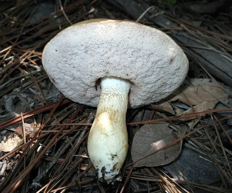
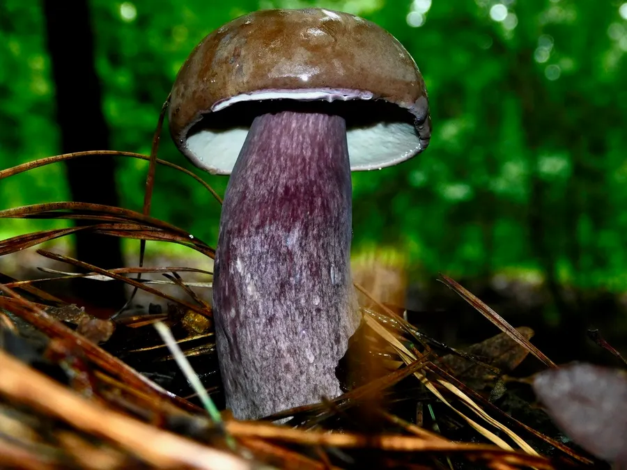
The cap of the fruit body is 7 to 15 cm in diameter, initially convex in shape but becoming centrally depressed, with a broadly arched and rounded margin. Young specimens are rather hard and firm, and the cap has a finely velvet-textured surface that soon wears off to become smooth. The color of the fruit body is violet when young, but dulls as it ages, becoming a dull violet-purplish-gray, then eventually chocolate-brown at maturity. The flesh is solid, white, and does not change color when cut or bruised. The taste is bitter, and the odor is not distinctive. Mycologist David Arora calls the mushroom "beautiful, but bitter-tasting". The tubes on the underside of the cap are 0.4 to 1.8 cm deep, 2 or three per millimeter, depressed at the stem (resulting in an adnate attachment). The color of the pore surface is initially white, and it remains so for a while before turning a rosy color at maturity. The stem is 8 to 13 cm long and 2.5 to 4 cm thick, enlarged at the base, and sometimes bulbous. The surface is slightly reticulate at the top, and smooth lower of the stem. Its color is buff to light brown, often with darker brown bruises or stains, and it has whitish mycelium at the base. The flesh of the stem is white, and it does not change color when cut or bruised. Collected in deposit, like with a spore print, the spores of T. plumbeoviolaceus appear to be a light pink to flesh color. When viewed with a light microscope, they are elliptical, with smooth walls and dimensions of 9.1–12.3 by 3.4–4.5 μm. The basidia (cellular structures that produce the spores) are club-shaped, and measure about 26 by 6.5 μm.
Habitat dan peran ekologis
Tylopilus plumbeoviolaceous adalah spesies mikoriza, dan timur Rocky Mountains from Canada to Meksiko. Spesies ini juga telah dikumpulkan di Korea Utara. Fruit bodies dapat berfungsi sebagai sumber makanan untuk pakan jamur Drosophila flies.
- Habitat atau konteks temuan
- Informasi belum tersedia pada sumber rilis ini.
- Substrat
- Informasi belum tersedia pada sumber rilis ini.
- Peran atau konteks
- Mitra tumbuhan
Reproduksi
Collected in deposit, like with a spore print, the spores of T. plumbeoviolaceus appear to be a light pink to flesh color. When viewed with a light microscope, they are elliptical, with smooth walls and dimensions of 9.1–12.3 by 3.4–4.5 μm. The basidia (cellular structures that produce the spores) are club-shaped, and measure about 26 by 6.5 μm. The cuticle of the cap (the pileipellis) is made of a tangle of smooth-walled, narrow, brownish hyphae. When stained in potassium hydroxide, the hyphal contents tend to form beads, while staining in Melzer's reagent causes the pigment to form globules. Cystidia are common in the hymenial tissue; they are swollen at the base and narrow at the apex (lageniform), measuring 30–40 μm long by 7–9 μm thick.
Catatan identitas dan cakupan
- Nama diterima mengikuti Catalogue of Life Base2026-09-11; uraian mempertahankan konsep dan nama sumber aslinya.
- Original geographic, specimen and life-cycle scope retained; source import awaits individual editorial review.
- Impor dan terjemahan otomatis belum diperiksa editorial per profil. Struktur reproduktif yang disebutkan tidak membuktikan seluruh siklus hidup diketahui.
- Pengukuran pada uraian mengikuti spesimen, kultur, dan metode sumber; angka tidak diperluas ke semua populasi.
Catatan distribusi dalam narasi mengikuti sumbernya. Dokumentasi spesimen atau foto mengikuti catatan sumber. Bukti wilayah Indonesia dan penilaian konservasi tidak disimpulkan dari gambar.
Melihat dengan konteks
Dokumentasi Tylopilus plumbeoviolaceus menurut asosiasi foto pada record takson iNaturalist. Kredit: Michael J. Papay. Identitas mengikuti sumber; bukan penentuan spesimen independen.
- Jenis
- Fotografi
- Kredit
- (c) Michael J. Papay,保留部分权利(CC BY), 由 Michael J. Papay 上传
- Lisensi
- CC BY 4.0
- Sumber media
- Buka entri sumber
Dokumentasi tambahan
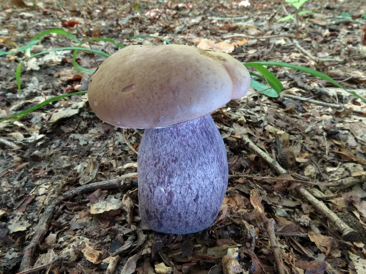
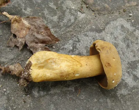
Foto yang diasosiasikan dengan Tylopilus plumbeoviolaceus pada iNaturalist. Identifikasi mengikuti sumber; bukan penentuan spesimen independen.
Kredit dan penggunaan
(c) Katja Schulz, some rights reserved (CC BY)
Ukuran dan format gambar disesuaikan tanpa pemotongan, retus, atau penambahan skala. Lisensi sumber dipertahankan.
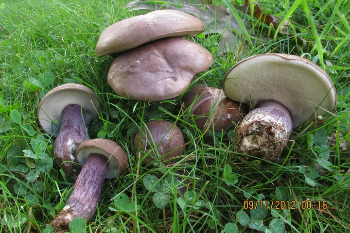
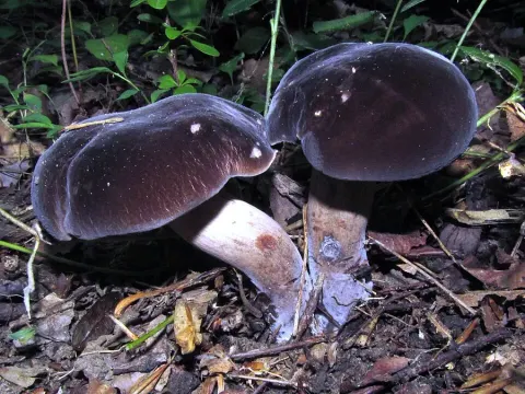
Foto yang diasosiasikan dengan Tylopilus plumbeoviolaceus pada iNaturalist. Identifikasi mengikuti sumber; bukan penentuan spesimen independen.
Kredit dan penggunaan
(c) mycowalt, some rights reserved (CC BY-SA), uploaded by mycowalt
Ukuran dan format gambar disesuaikan tanpa pemotongan, retus, atau penambahan skala. Lisensi sumber dipertahankan.
Sumber profil
- [1] Tylopilus plumbeoviolaceus — Wikipedia (en)
- [2] Catalogue of Life Base2026-09-11 — Tylopilus plumbeoviolaceus
- [3] Dokumentasi visual — Tylopilus plumbeoviolaceus
- [4] Dokumentasi visual — Tylopilus plumbeoviolaceus
- [5] Dokumentasi visual — Tylopilus plumbeoviolaceus
Rujukan halaman ini
Fungi. Tylopilus plumbeoviolaceus. Rilis data fungi-2026-10-12-expansion-2. https://fungi-atlas.mgadib.chatgpt.site/jamur/tylopilus-plumbeoviolaceus/.
Status: impor sumber; pemeriksaan editorial per profil belum selesai. Untuk klaim biologis, gunakan rujukan pada bagian terkait dan periksa konteks sumber aslinya.