Ciri dan struktur
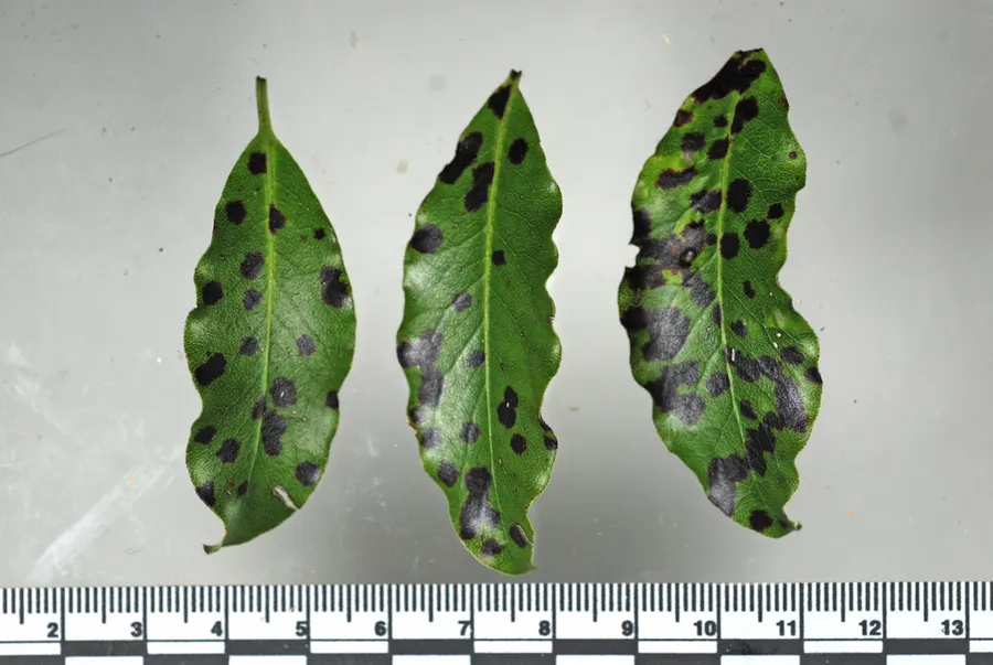
Leaf spots on the upper surface of the leaf, roughly circular, 2-12 mm diameter, dull black, necrotic, mostly discrete but sometimes confluent. Mycelium subcuticular, with light-brown and hyaline cells in no particular order, hyphae 5-7 µm wide with individual cells varying from 5 to 35 µm in length, septa thickened and dark-brown. Ascocarp a thyrothecium, fruitbodies arranged in groups on the black spots, discrete, shining black, dimidate or flattened convex, 120-160 µm in diameter. The ascocarp has an ostiole, 10-20 µm diameter, at maturity. Upper wall of the ascocarp composed of two layers of dark-brown, non-radiating cells, 3-5 µm wide. Lower wall composed of a single layer of subhyaline cells, 3-5 µm wide. Asci arranged radially round a central column of paraphysoid tissue, bitunicate, ellipsoidal to subclavate, 45-65 µm long x 15-20 µm wide, with 8 spores, apex rounded and thickened. Ascospores arranged in 1-2 rows, unicellular, obovate, brown with a transverse hyaline zone (1-2 µm wide) just above the middle, 10-14 µm long x 5-7 µm wide, with a small (about 2 µm long), conical, hyaline basal appendage. A thin gelatinous coat of uniform thickness surrounds the ascospore. Conidia borne in structures similar to ascocarps, ellipsoidal, hyaline, 5-8 x 4-6 µm. Conidiophores not seen.
Habitat dan peran ekologis
On living leaves of Pittosporum tenuifolium and of Myrsine australis. The specific epithet refers to the gelatinous coat surrounding the ascospore. The main characteristic that distinguishes the New Zealand species from other Vizella spp. is the gelatinous coat surrounding each ascospore. Müller & von Arx (1955), after examining Vizella gomphispora (Berkeley & Broome) Hughes, V. royenae von Arx & Müller, and V. bingervilliana C.Moreau & M.Moreau, concluded that these three species were conspecific with the older V. appendiculosa (Montagne & Berkeley) Theissen. In addition to the possession of a gelatinous coat, the New Zealand species differs from V. appendiculosa sensu lato in that (a) its ascospores have the hyaline band in the middle rather than close to the apex (Hughes 1953; von Arx & Müller 1975) and (b) rather than having thick hyphae composed of "almost cuboid" cells up to 12 µm wide (Hughes 1953), the width of its hyphal cells does not exceed 7 µm and the length varies from 5 µm to 35 µm. In common with the Australian V. oleariae Swart, both the conidial and ascal states of the New Zealand species are found in the same colony. However, the conidia of the New Zealand species, unlike those of V. oleariae, remain hyaline at maturity and the mycelium does not have the regular alternation of paired hyaline cells as with dark cells as described for V. oleariae (Swart 1971).
- Habitat atau konteks temuan
- Informasi belum tersedia pada sumber rilis ini.
- Substrat
- Informasi belum tersedia pada sumber rilis ini.
- Peran atau konteks
- Belum dikategorikan pada rilis ini.
Reproduksi
Leaf spots on the upper surface of the leaf, roughly circular, 2-12 mm diameter, dull black, necrotic, mostly discrete but sometimes confluent. Mycelium subcuticular, with light-brown and hyaline cells in no particular order, hyphae 5-7 µm wide with individual cells varying from 5 to 35 µm in length, septa thickened and dark-brown. Ascocarp a thyrothecium, fruitbodies arranged in groups on the black spots, discrete, shining black, dimidate or flattened convex, 120-160 µm in diameter. The ascocarp has an ostiole, 10-20 µm diameter, at maturity. Upper wall of the ascocarp composed of two layers of dark-brown, non-radiating cells, 3-5 µm wide. Lower wall composed of a single layer of subhyaline cells, 3-5 µm wide.
Catatan identitas dan cakupan
- Nama diterima mengikuti Catalogue of Life Base2026-09-11; uraian mempertahankan konsep dan nama sumber aslinya.
- Historical description as cited by BiotaNZ. Preserve explicit New Zealand/locality, host and specimen qualifiers. Distribution is the cited source scope, not a current global range.
- Impor dan terjemahan otomatis belum diperiksa editorial per profil. Struktur reproduktif yang disebutkan tidak membuktikan seluruh siklus hidup diketahui.
- Pengukuran pada uraian mengikuti spesimen, kultur, dan metode sumber; angka tidak diperluas ke semua populasi.
Catatan distribusi dalam narasi mengikuti sumbernya. Dokumentasi spesimen atau foto mengikuti catatan sumber. Bukti wilayah Indonesia dan penilaian konservasi tidak disimpulkan dari gambar.
Melihat dengan konteks
Dokumentasi Vizella tunicata menurut asosiasi foto pada record takson iNaturalist. Kredit: Jerry Cooper. Identitas mengikuti sumber; bukan penentuan spesimen independen.
- Jenis
- Fotografi
- Kredit
- (c) Jerry Cooper, some rights reserved (CC BY), uploaded by Jerry Cooper
- Lisensi
- CC BY 4.0
- Sumber media
- Buka entri sumber
Dokumentasi tambahan
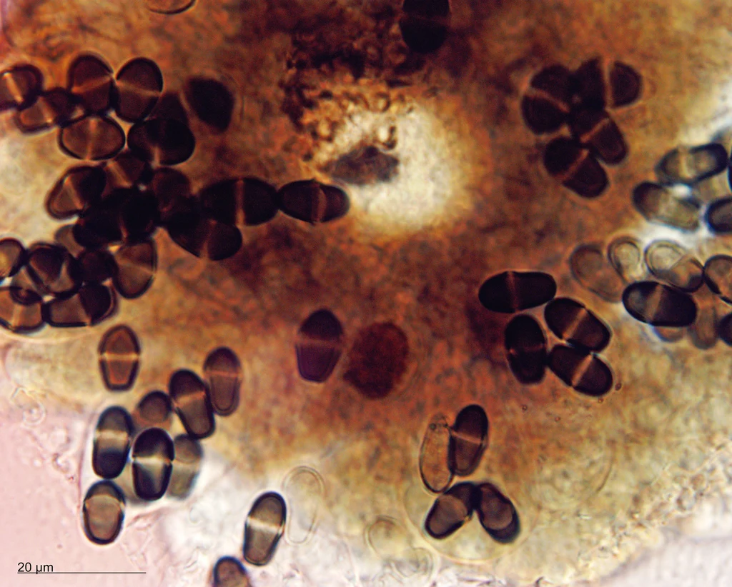
Preparat mikroskopis yang diasosiasikan dengan Vizella tunicata pada iNaturalist. Identitas mengikuti sumber.
Kredit dan penggunaan
(c) Jerry Cooper, some rights reserved (CC BY), uploaded by Jerry Cooper
Ukuran dan format gambar disesuaikan tanpa pemotongan, retus, atau penambahan skala. Lisensi sumber dipertahankan.
Sumber profil
- [1] Gadgil, P.D. 1995: Mycological records 4: Vizella tunicata sp. nov. New Zealand Journal of Forestry Science 25(1): 107-110. — Vizella tunicata
- [2] Catalogue of Life Base2026-09-11 — Vizella tunicata
- [3] Dokumentasi visual — Vizella tunicata
- [4] Dokumentasi visual — Vizella tunicata
Rujukan halaman ini
Fungi. Vizella tunicata. Rilis data fungi-2026-10-12-expansion-2. https://fungi-atlas.mgadib.chatgpt.site/jamur/vizella-tunicata/.
Status: impor sumber; pemeriksaan editorial per profil belum selesai. Untuk klaim biologis, gunakan rujukan pada bagian terkait dan periksa konteks sumber aslinya.