Ciri dan struktur
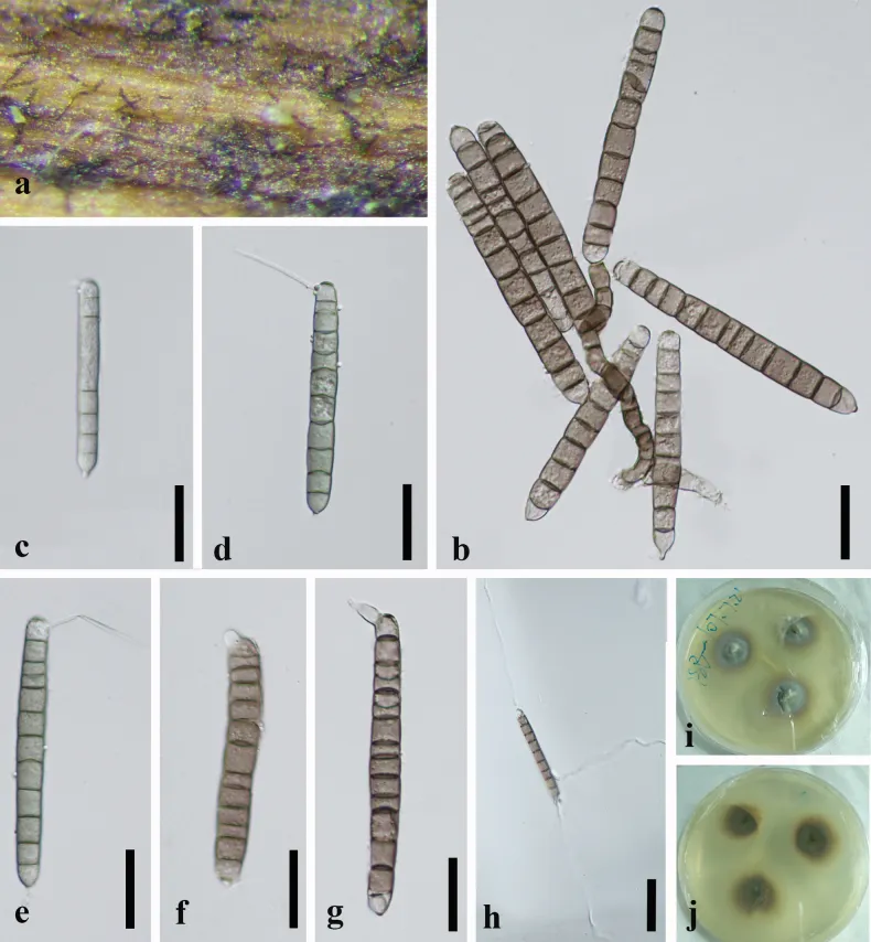
Saprobic on decaying, submerged wood in a freshwater habitat. Asexual morph. Colonies on natural substrate effuse, brown, procumbent. Mycelium partly immersed and partly superficial, composed of pale brown, septate, cylindrical hyphae. Conidiophores macronematous, mononematous, irregularly cylindrical, strongly flexuous to twisted, procumbent, brown, sometimes fading slightly towards the apex, smooth-walled, unbranched, thick-walled, 4–8–septate, 75–92 × 5.5–6.5 μm (x̄= 86 × 6 μm, n = 20). Conidiogenous cell: holoblastic, monoblastic, integrated, terminal, cylindrical, pale brown. Conidia solitary, cylindrical to narrowly fusoid, elongate, subhyaline to pale olivaceous green when young, brown or pale brown when mature, polar cells slightly paler, 64–114 × 7.5–11.5 μm (x̄= 91.5 × 9.5 μm, n = 30), smooth-walled, verruculose, 6–13-septate, often slightly constricted at the septa, basal cell conical, apical cell rounded with 2 independent, simple, cellular, aseptate, hyaline, smooth, straight, curved, or flexuous appendages, 29.5–40 × 1.5–2.5 μm (x̄= 33 × 1.8 μm, n = 20). Sexual morph. Undetermined. Conidia germinated on WA within 24 h, and germ tubes were produced from the apex. The colonies on PDA reached approximately 22 mm diam. after 3 weeks at 25 °C under dark conditions. Colonies circular, with raised center and filamentous edge, grey at center, brown at the edge from above, dark brown in the center, brown at the edge from reverse.
Habitat dan peran ekologis
Saprobic on decaying, submerged wood in a freshwater habitat. Asexual morph. Colonies on natural substrate effuse, brown, procumbent. Mycelium partly immersed and partly superficial, composed of pale brown, septate, cylindrical hyphae. Conidiophores macronematous, mononematous, irregularly cylindrical, strongly flexuous to twisted, procumbent, brown, sometimes fading slightly towards the apex, smooth-walled, unbranched, thick-walled, 4–8–septate, 75–92 × 5.5–6.5 μm (x̄= 86 × 6 μm, n = 20). Conidiogenous cell: holoblastic, monoblastic, integrated, terminal, cylindrical, pale brown. Conidia solitary, cylindrical to narrowly fusoid, elongate, subhyaline to pale olivaceous green when young, brown or pale brown when mature, polar cells slightly paler, 64–114 × 7.5–11.5 μm (x̄= 91.5 × 9.5 μm, n = 30), smooth-walled, verruculose, 6–13-septate, often slightly constricted at the septa, basal cell conical, apical cell rounded with 2 independent, simple, cellular, aseptate, hyaline, smooth, straight, curved, or flexuous appendages, 29.5–40 × 1.5–2.5 μm (x̄= 33 × 1.8 μm, n = 20). Sexual morph. Undetermined. Conidia germinated on WA within 24 h, and germ tubes were produced from the apex. The colonies on PDA reached approximately 22 mm diam. after 3 weeks at 25 °C under dark conditions.
- Habitat atau konteks temuan
- kultur yang didokumentasikan
- Substrat
- Informasi belum tersedia pada sumber rilis ini.
- Peran atau konteks
- Pengurai
Reproduksi
Saprobic on decaying, submerged wood in a freshwater habitat. Asexual morph. Colonies on natural substrate effuse, brown, procumbent. Mycelium partly immersed and partly superficial, composed of pale brown, septate, cylindrical hyphae. Conidiophores macronematous, mononematous, irregularly cylindrical, strongly flexuous to twisted, procumbent, brown, sometimes fading slightly towards the apex, smooth-walled, unbranched, thick-walled, 4–8–septate, 75–92 × 5.5–6.5 μm (x̄= 86 × 6 μm, n = 20). Conidiogenous cell: holoblastic, monoblastic, integrated, terminal, cylindrical, pale brown. Conidia solitary, cylindrical to narrowly fusoid, elongate, subhyaline to pale olivaceous green when young, brown or pale brown when mature, polar cells slightly paler, 64–114 × 7.5–11.5 μm (x̄= 91.5 × 9.5 μm, n = 30), smooth-walled, verruculose, 6–13-septate, often slightly constricted at the septa, basal cell conical, apical cell rounded with 2 independent, simple, cellular, aseptate, hyaline, smooth, straight, curved, or flexuous appendages, 29.5–40 × 1.5–2.5 μm (x̄= 33 × 1.8 μm, n = 20).
Catatan identitas dan cakupan
- Nama diterima mengikuti Catalogue of Life Base2026-09-11; uraian mempertahankan konsep dan nama sumber aslinya.
- Preserve original treatment localities, hosts and substrate scope. Materials examined describe documented specimens, not the full global range.
- Impor dan terjemahan otomatis belum diperiksa editorial per profil. Struktur reproduktif yang disebutkan tidak membuktikan seluruh siklus hidup diketahui.
- Pengukuran pada uraian mengikuti spesimen, kultur, dan metode sumber; angka tidak diperluas ke semua populasi.
Catatan distribusi dalam narasi mengikuti sumbernya. Dokumentasi spesimen atau foto mengikuti catatan sumber. Bukti wilayah Indonesia dan penilaian konservasi tidak disimpulkan dari gambar.
Melihat dengan konteks
Camposporium aquaticum (GZAAS 20-0376, Herbarium) a colonies on natural substrate b conidiophore and conidium c–g conidia h germinating conidia i, j culture i from above j from below. Scale bars: 25 μm (c–j); 100 μm (h).
- Jenis
- Pelat ilmiah
- Kredit
- Ling-Ling Liu, Yong-Xiang Liu, Ya-Ya Chen, Jiu-Lan Gou, Feng Chi, Yi Liu, Xiao-Feng Gu, Quan-Quan Wei, Meng Zhang, Zuo-Yi Liu, Si Zhou
- Lisensi
- CC BY 4.0
- Sumber media
- Buka entri sumber
Sumber profil
- [1] Freshwater fungi in the karst plateau wetlands from Guizhou Province, China: taxonomic novelties in Melanommataceae (Pleosporales) — Camposporium aquaticum
- [2] Catalogue of Life Base2026-09-11 — Xylomyces aquaticus
- [3] Dokumentasi visual — Camposporium aquaticum
Rujukan halaman ini
Fungi. Xylomyces aquaticus. Rilis data fungi-2026-10-12-expansion-2. https://fungi-atlas.mgadib.chatgpt.site/jamur/xylomyces-aquaticus/.
Status: impor sumber; pemeriksaan editorial per profil belum selesai. Untuk klaim biologis, gunakan rujukan pada bagian terkait dan periksa konteks sumber aslinya.